Molecular-level secondary structure, polymorphism, and dynamics of full-length alpha-synuclein fibrils studied by solid-state NMR
- PMID: 16247008
- PMCID: PMC1276071
- DOI: 10.1073/pnas.0506109102
Molecular-level secondary structure, polymorphism, and dynamics of full-length alpha-synuclein fibrils studied by solid-state NMR
Abstract
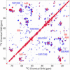
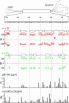
The 140-residue protein alpha-synuclein (AS) is able to form amyloid fibrils and as such is the main component of protein inclusions involved in Parkinson's disease. We have investigated the structure and dynamics of full-length AS fibrils by high-resolution solid-state NMR spectroscopy. Homonuclear and heteronuclear 2D and 3D spectra of fibrils grown from uniformly (13)C/(15)N-labeled AS and AS reverse-labeled for two of the most abundant amino acids, K and V, were analyzed. (13)C and (15)N signals exhibited linewidths of <0.7 ppm. Sequential assignments were obtained for 48 residues in the hydrophobic core region. We identified two different types of fibrils displaying chemical-shift differences of up to 13 ppm in the (15)N dimension and up to 5 ppm for backbone and side-chain (13)C chemical shifts. EM studies suggested that molecular structure is correlated with fibril morphology. Investigation of the secondary structure revealed that most amino acids of the core region belong to beta-strands with similar torsion angles in both conformations. Selection of regions with different mobility indicated the existence of monomers in the sample and allowed the identification of mobile segments of the protein within the fibril in the presence of monomeric protein. At least 35 C-terminal residues were mobile and lacked a defined secondary structure, whereas the N terminus was rigid starting from residue 22. Our findings agree well with the overall picture obtained with other methods and provide insight into the amyloid fibril structure and dynamics with residue-specific resolution.
Figures


Similar articles
-
Structural comparison of mouse and human α-synuclein amyloid fibrils by solid-state NMR.J Mol Biol. 2012 Jun 29;420(1-2):99-111. doi: 10.1016/j.jmb.2012.04.009. Epub 2012 Apr 16. J Mol Biol. 2012. PMID: 22516611
-
Amyloid fibril formation by A beta 16-22, a seven-residue fragment of the Alzheimer's beta-amyloid peptide, and structural characterization by solid state NMR.Biochemistry. 2000 Nov 14;39(45):13748-59. doi: 10.1021/bi0011330. Biochemistry. 2000. PMID: 11076514
-
Solid-state NMR reveals structural differences between fibrils of wild-type and disease-related A53T mutant alpha-synuclein.J Mol Biol. 2008 Jul 11;380(3):444-50. doi: 10.1016/j.jmb.2008.05.026. Epub 2008 May 17. J Mol Biol. 2008. PMID: 18539297
-
Molecular structure of amyloid fibrils: insights from solid-state NMR.Q Rev Biophys. 2006 Feb;39(1):1-55. doi: 10.1017/S0033583506004173. Epub 2006 Jun 13. Q Rev Biophys. 2006. PMID: 16772049 Review.
-
The structure of fibrils from 'misfolded' proteins.Curr Opin Struct Biol. 2015 Feb;30:43-49. doi: 10.1016/j.sbi.2014.12.001. Epub 2014 Dec 24. Curr Opin Struct Biol. 2015. PMID: 25544255 Review.
Cited by
-
High resolution structural characterization of Aβ42 amyloid fibrils by magic angle spinning NMR.J Am Chem Soc. 2015 Jun 17;137(23):7509-18. doi: 10.1021/jacs.5b03997. Epub 2015 Jun 4. J Am Chem Soc. 2015. PMID: 26001057 Free PMC article.
-
How important is the N-terminal acetylation of alpha-synuclein for its function and aggregation into amyloids?Front Neurosci. 2022 Nov 16;16:1003997. doi: 10.3389/fnins.2022.1003997. eCollection 2022. Front Neurosci. 2022. PMID: 36466161 Free PMC article. Review.
-
Amyloid fibril formation kinetics of low-pH denatured bovine PI3K-SH3 monitored by three different NMR techniques.Front Mol Biosci. 2023 Nov 17;10:1254721. doi: 10.3389/fmolb.2023.1254721. eCollection 2023. Front Mol Biosci. 2023. PMID: 38046811 Free PMC article.
-
Chicago sky blue 6B inhibits α-synuclein aggregation and propagation.Mol Brain. 2022 Mar 28;15(1):27. doi: 10.1186/s13041-022-00913-y. Mol Brain. 2022. PMID: 35346306 Free PMC article.
-
Atomic resolution protein structure determination by three-dimensional transferred echo double resonance solid-state nuclear magnetic resonance spectroscopy.J Chem Phys. 2009 Sep 7;131(9):095101. doi: 10.1063/1.3211103. J Chem Phys. 2009. PMID: 19739873 Free PMC article.
References
Publication types
MeSH terms
Substances
LinkOut - more resources
Full Text Sources
Other Literature Sources
Research Materials

