. 2005 Nov 1;102(44):16013-8.
doi: 10.1073/pnas.0500090102. Epub 2005 Oct 24.

The response of autologous T cells to a human melanoma is dominated by mutated neoantigens

Affiliations

The response of autologous T cells to a human melanoma is dominated by mutated neoantigens

Volker Lennerz et al. Proc Natl Acad Sci U S A. .

Abstract

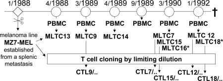
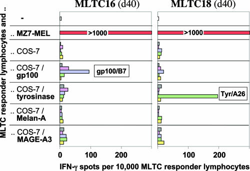
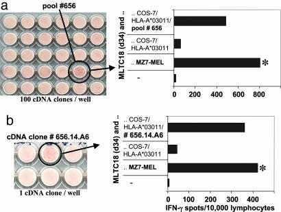
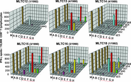
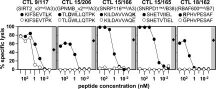
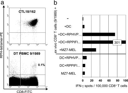
Our understanding of pathways leading to antitumor immunity may depend on an undistorted knowledge of the primary antigenic targets of patients' autologous T cell responses. In the melanoma model derived from patient DT, we applied cryopreserved short-term autologous mixed lymphocyte-tumor cell cultures (MLTCs) in combination with an IFN-gamma enzyme-linked immunospot (ELISPOT) assay to cDNA expression screening. We identified three previously unknown peptides processed from melanosomal proteins tyrosinase (presented by HLA-A(*)2601 and -B(*)3801) and gp100 (presented by HLA-B(*)07021) and five neoantigens generated by somatic point mutations in the patient's melanoma. The mutations were found in the genes SIRT2, GPNMB, SNRP116, SNRPD1, and RBAF600. Peptides containing the mutated residues were presented by HLA-A(*)03011, -B(*)07021, and -B(*)3801. Mutation-induced functional impairment was so far demonstrated for SIRT2. Within MLTC responder populations that were independently expanded from the patient's peripheral blood lymphocytes of different years, T cells against mutated epitopes clearly predominated. These results document a high degree of individuality for the cellular antitumor response and support the need for individualizing the monitoring and therapeutic approaches to the primary targets of the autologous T cell response, which may finally lead to a more effective cancer immunotherapy.


Figures

Fig. 1.
Fig. 1.
Origin of MLTC and CTL clones. MLTC were performed by stimulating PBMC taken from patient DT at indicated time points (month/year) with autologous tumor cells (MZ7-MEL) as detailed in Materials and Methods. CTL clones were derived by limiting dilution from some of the MLTC (designation x/y for CTL clones: x was the serial number of the MLTC, y was the serial number of the T cell clone). *, Stimulator cells were pretreated with IFN-γ.
Fig. 2.
Fig. 2.
Reactivity of MLTC responders to known common antigens. Responder lymphocytes of MLTC16(d40) and MLTC18(d40) (10,000 lymphocytes per well) were tested in IFN-γ ELISPOT assays for recognition of antigens MAGE-A3, tyrosinase, gp100, and Melan-A/MART-1. Data are means of duplicates. Stimulators were melanoma cells (MZ7-MEL, 30,000 per well) and COS-7 cells (20,000 per well) cotransfected with antigen-coding cDNA (indicated) and HLA class I cDNA cloned by RT-PCR from MZ7-MEL cells: yellow, HLA-A*03011; green, HLA-A*2601; purple, HLA-B*07021; pink, HLA-B*3801; gray, HLA-Cw*1203.
Fig. 3.
Fig. 3.
Cloning of antigen-coding cDNA with cryopreserved MLTC18 responders. MLTC18(d25) contained T cells against Tyr/A26 at low frequencies, but was strongly reactive with autologous melanoma cells (Fig. 2). Its antitumor reactivity was partially inhibited by an anti-HLA-A3 monoclonal antibody (data not shown). MLTC18 was further expanded. On day 32, 2.78 × 10⁸ CD8+ T cells were frozen in aliquots 4 days after the last stimulation with autologous tumor cells. For cDNA library screening, aliquots were thawed and T cells were kept for 2 days in medium with IL-2. In parallel, COS-7 cells were cotransfected directly in MultiScreen ELISPOT HP plates with pools of ≈100 cDNA clones and with HLA-A*03011 cDNA. After 24 h, MLTC responders were added at 10,000 lymphocytes per well. Each cDNA pool was then tested for its ability to induce IFN-γ spot formation. Thirteen of 1,920 cDNA pools were positive. A filter section with positive pool 656 is shown next to the spot evaluation data (a). From pool 656, we identified cDNA clone 656.14.A6 as inducer of spot formation after cotransfection with HLA-A*03011 (b). This cDNA clone encoded GPNMB_v2mut (Table 1). Only 11 of 13 positive “pools of 100” identified in this screening experiment contained GPNMB_v2mut cDNA as verified by PCR. From the two remaining pools, we isolated a cDNA clone encoding SNRP116mut (Table 1). *, Reactivity to melanoma cells was tested with the same MLTC population in parallel (30,000 MZ7-MEL cells per well; a, 1,000 lymphocytes per well; b, 500 lymphocytes per well).
Fig. 4.
Fig. 4.
Specificity of tumor-reactive T cells enriched in DT MLTC. Independent autologous MLTC were performed with PBMC collected from patient DT during a 4-year period (Fig. 1). CD8+ MLTC responders were isolated and cryopreserved at various time points and later tested in 20-h IFN-γ ELISPOT assays for recognition of autologous melanoma cells (30,000 per well) (M) and COS-7 cells (20,000 per well) transiently transfected with expression plasmids encoding the antigen/HLA combinations tyrosinase/HLA-A*2601 (A), tyrosinase/HLA-B*3801 (B), gp100/HLA-B*07021 (C), SIRT2_v3mut/HLA-A*03011 (D), GPNMB_v2mut/HLA-A*03011 (E), SNRP116mut/HLA-A*03011 (F), RBAF600mut/HLA-B*07021 (G), SNRPD1mut/HLA-B*3801 (H). Data are means of duplicates and represent the number of spot forming cells per 15,000 (day 25) or 10,000 (later than day 25) CD8+ MLTC responders.
Fig. 5.
Fig. 5.
Titration of peptides. Data of a 4-h 51Cr release assay are shown. Peptides (sequences and concentrations as indicated, mutated residues bold and underlined, see Table 1) were directly added to 51Cr-labeled MZ7-EBV-B cells (2,000 per well). CTL clones (specificity indicated in parentheses) were added at an effector-to-target (E/T) cell ratio of 20:1. Filled circles, mutated peptides; open circles, homologous wild-type peptides; filled diamonds, lysis of MZ7-MEL cells (E/T = 20:1, 2,000 melanoma cells per well).
Fig. 6.
Fig. 6.
Detection of RBAF600mut/B7-reactive T cells in ex vivo PBMC. (a) PBMCs taken from patient DT in September 1989 and, as a control, the RBAF600mut/B7-specific T cell clone CTL18/162, were stained with phycoerythrin (PE)-conjugated tetrameric complex B7/RPHVPESAF containing the mutant RBAF600 peptide and HLA-B7 and with an antibody to CD8 conjugated to FITC. (b) In parallel, CD8+ T cells were positively selected from the same PBMC and were analyzed by a 48-h IFN-γ ELISPOT assay using peptide-pulsed autologous DCs (20,000 per well) and melanoma cells (30,000 per well) as antigen-presenting cells. Peptides were the RBAF600mut/B7 peptide RPH-VPESAF (Table 1) and, as a positive control, the HLA-B7-restricted EBNA 3A 379-387 peptide RPPIFIRRL (14).

References

    1. Van den Eynde, B. J. & van der Bruggen, P. (1997) Curr. Opin. Immunol. 9, 684-693. - PubMed
    1. Rosenberg, S. A. (1999) Immunity 10, 281-287. - PubMed
    1. Srivastava, P. J. & Old, L. J. (1988) Immunol. Today 9, 78-83. - PubMed
    1. Beck-Engeser, G. B., Monach, P. A., Mumberg, D., Yang, F., Wanderling, S., Schreiber, K., Espinosa, R., III, Le Beau, M. M., Meredith, S. C. & Schreiber, H. (2001) J. Exp. Med. 194, 285-300. - PMC - PubMed
    1. Gilboa, E. (1999) Immunity 11, 263-270. - PubMed

Publication types