. 2005 Nov 1;102(44):15901-6.
doi: 10.1073/pnas.0508014102. Epub 2005 Oct 24.

Metastatic properties and genomic amplification of the tyrosine kinase gene ACK1

Affiliations

Metastatic properties and genomic amplification of the tyrosine kinase gene ACK1

Edward Htun van der Horst et al. Proc Natl Acad Sci U S A. .

Abstract

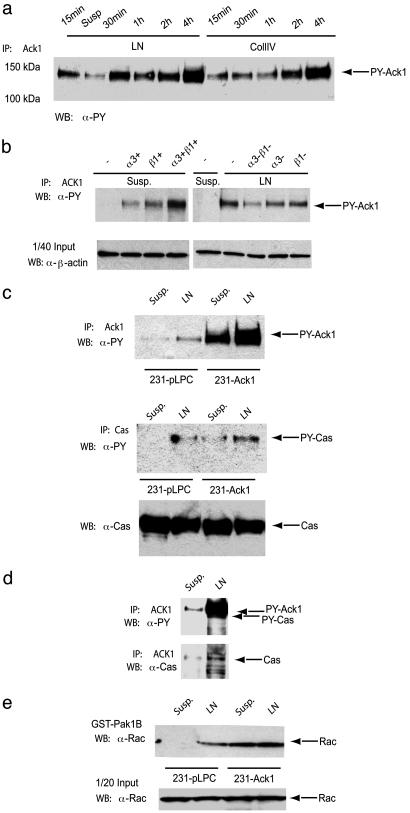
Metastasis of primary tumors leads to a very poor prognosis for patients suffering from cancer. Although it is well established that not every tumor will eventually metastasize, it is less clear whether primary tumors acquire genetic alterations in a stochastic process at a late stage, which make them invasive, or whether genetic alterations acquired early in the process of tumor development drive primary tumor growth and determine whether this tumor is going to be metastatic. To address this issue, we tested genes identified in a large-scale comparative genomic hybridization analysis of primary tumor for their ability to confer metastatic properties on a cancer cell. We identified amplification of the ACK1 gene in primary tumors, which correlates with poor prognosis. We further show that overexpression of Ack1 in cancer cell lines can increase the invasive phenotype of these cells both in vitro and in vivo and leads to increased mortality in a mouse model of metastasis. Biochemical studies show that Ack1 is involved in extracellular matrix-induced integrin signaling, ultimately activating signaling processes like the activation of the small GTPase Rac. Taken together, this study supports a theory from Bernards and Weinberg [Bernards, R. & Weinberg, R. A. (2002) Nature 418, 823], which postulates that the tendency to metastasize is largely predetermined.


Figures

Fig. 1.
Fig. 1.
ACK1 is amplified in chromosome 3, overexpressed, and associated with poor prognosis in human tumors. (a) Genomic microarray analysis of relative DNA copy number along chromosome (Chr.) 3 in several tumor types. (b) DNA copy number for eight tumors (two hormone-refractory prostate tumors, red and blue asterisks; two ovarian tumors, solid blue and green quadrilaterals; two esophageal tumors, blue and red dashes; a lung tumor, solid green circles; and a pancreatic tumor, open magenta circles). DNA copy number is plotted in megabase pairs against their nucleotide position in chromosome 3 (http://genome.ucsc.edu; April 2003 freeze). The data shown are from single DNA TaqMan assays [each with duplicate measurements with coefficient of variation (CV) <5%], and each assay was performed three times with an average CV ≅ 10%. (c) Corresponding ACK1 DNA and RNA values for lung, ovarian, and prostate tumors as determined by QPCR, with relative RNA levels on a log scale. Ack1 RNA was measured by quantitative QPCR in normal prostate tissues, primary prostate tumors, and hormone-refractory prostate tumors (d) (values >50 times are shown as 50); and normal lung tissues, stage 1 and 2 lung tumors, and stage 3 and 4 tumors (e). The values were normalized to the average value obtained with five corresponding normal tissue samples.
Fig. 2.
Fig. 2.
Ack1 overexpression in tumor cells enhances metastasis and mortality in vivo.(a) Experimental metastasis assay with 231-pLPC and 231-Ack1 cells after 60 days. Human cells in mouse lungs were quantified by applying ALU-QPCR as described in ref. . Samples have been analyzed in quadruplicate and normalized against normal lung; each bar represents the results from an individual lung. (b and c) Colony formation of 4T1 cells isolated from lungs (b) or blood (c) of BALB/c mice 28 days after implantation. Tumor cells were cultivated for 21 days in vitro under selective conditions before colonies were fixed, stained, and counted. The experiment was performed twice with 10 and 30 mice per group (data shown are average of 30 individual samples). (d) Kaplan–Meier survival analysis of BALB/c mice inoculated with 4T1 cells overexpressing Ack1 or vector control. The primary tumor was removed when it reached a volume of 1,000–1,500 mm3, and the animals were monitored until they were moribund (44 animals per group). The experiment was stopped when all animals in the Ack1 group died and statistical significance was reached (P < 0.0001; paired t test).
Fig. 3.
Fig. 3.
Ack1 overexpression enhances invasiveness in vitro, phenotypical changes, and enhanced motility in HMEC and 4T1 cells. (a) HMEC-pLPC or HMEC-Ack1 cells were plated at low-density and photographed after 3 days. A representative photograph is shown. (b) Expression pattern of epithelial-marker (Left), mesenchymal-marker proteins (Right), and Ack1 expression in HMEC-pLPC and HMEC-Ack1 cells. (c) Matrigel-invasion assay with HMEC-pLPC or HMEC-Ack1 and 4T1-pLPC or 4T1-Ack1 cells after 24 h. The experiment was performed twice in octaduplicates (P < 0.001; Student's t test). Invasiveness has been quantified as described in the legend of Fig. 5.
Fig. 4.
Fig. 4.
Ack1 is tyrosine-phosphorylated upon α3β1-ligation, associates with p130Cas, and enhances GTP loading of Rac in the breast cancer cell line MDA-MB-231. (a) Tyrosine phosphorylation of Ack1 displays different kinetics when plated onto laminin (LN) or collagen IV (CollIV) plates in MDA-MB-231 cells. Please note that in Fig. 1a, lane 2 corresponds to cells kept in suspension. (b) Tyrosine phosphorylation of Ack1 depends on α3β1-integrin ligation. MDA-MB-231 cells were pretreated for 30 min with either stimulatory (+)- or inhibitory (-)-integrin antibodies before plating onto LN or plastic (PL). (c) Forced expression of Ack1 increases tyrosine phosphorylation of p130Cas upon LN stimulation in 231-Ack1 cells. (d) Ack1 associates with p130Cas upon LN stimulation in parental MDA-MB-231 cells. (e) GST-Pak1B pull down with 231-pLPC and 231-Ack1 cells after laminin stimulation. Cell lysates were subjected to immunoprecipitation (IP) with anti-Ack1 (Ack1) (a–d topmost panel) or p130Cas (c Middle). Tyrosine phosphorylation was analyzed by Western blotting (WB) with monoclonal antiphosphotyrosine antibody (α-PY). Loading of proteins and coprecipitation of p130Cas was controlled by reblotting with α-Ack1 and α-p130Cas. GTP-loaded Rac was analyzed by Western blotting with anti-Rac antibody (α-Rac). Arrows indicate the detected proteins.

References

    1. Fidler, I. J. & Kripke, M. L. (1977) Science 197, 893-895. - PubMed
    1. Poste, G. & Fidler, I. J. (1980) Nature 283, 139-146. - PubMed
    1. Israeli, O., Gotlieb, W. H., Friedman, E., Korach, J., Goldman, B., Zeltser, A., Ben-Baruch, G., Rienstein, S. & Aviram-Goldring, A. (2004) Cancer Genet. Cytogenet. 154, 16-21. - PubMed
    1. Patmore, H. S., Ashman, J. N., Cawkwell, L., MacDonald, A., Stafford, N. D. & Greenman, J. (2004) Br. J. Cancer 90, 1976-1982. - PMC - PubMed
    1. van 't Veer, L. J., Dai, H., van de Vijver, M. J., He, Y. D., Hart, A. A., Bernards, R. & Friend, S. H. (2003) Breast Cancer Res. 5, 57-58. - PMC - PubMed

MeSH terms