. 2005 Oct 24;171(2):267-79.
doi: 10.1083/jcb.200506124.

A complex containing the Sm protein CAR-1 and the RNA helicase CGH-1 is required for embryonic cytokinesis in Caenorhabditis elegans

Affiliations

A complex containing the Sm protein CAR-1 and the RNA helicase CGH-1 is required for embryonic cytokinesis in Caenorhabditis elegans

Anjon Audhya et al. J Cell Biol. .

Abstract

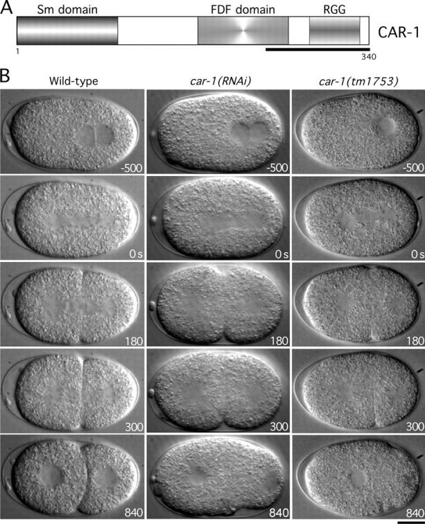
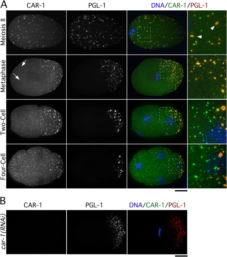
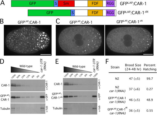
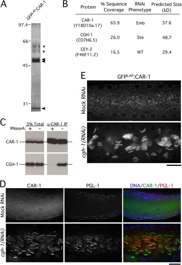
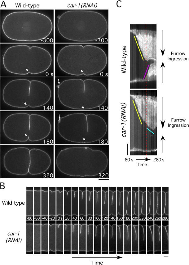
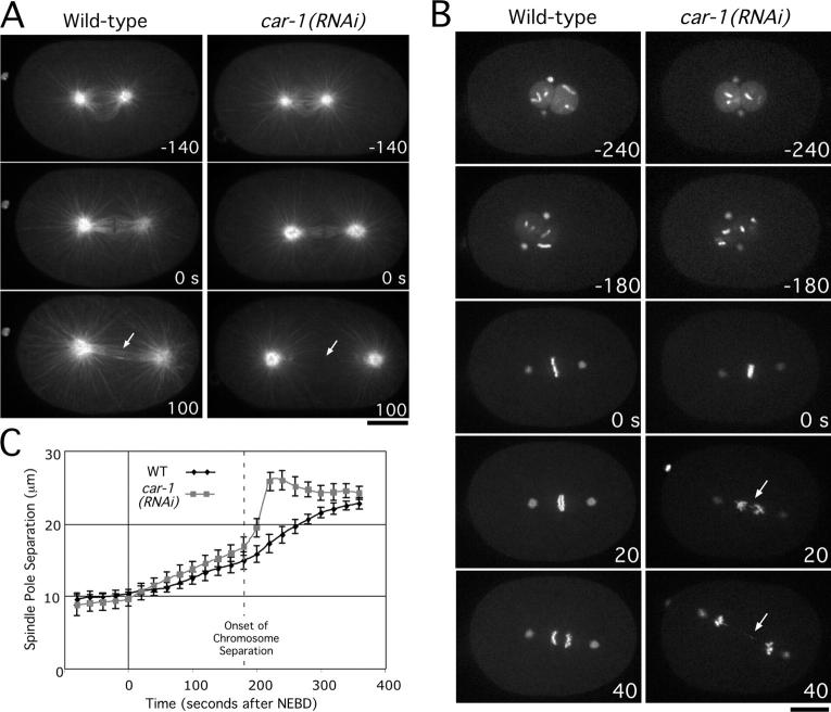
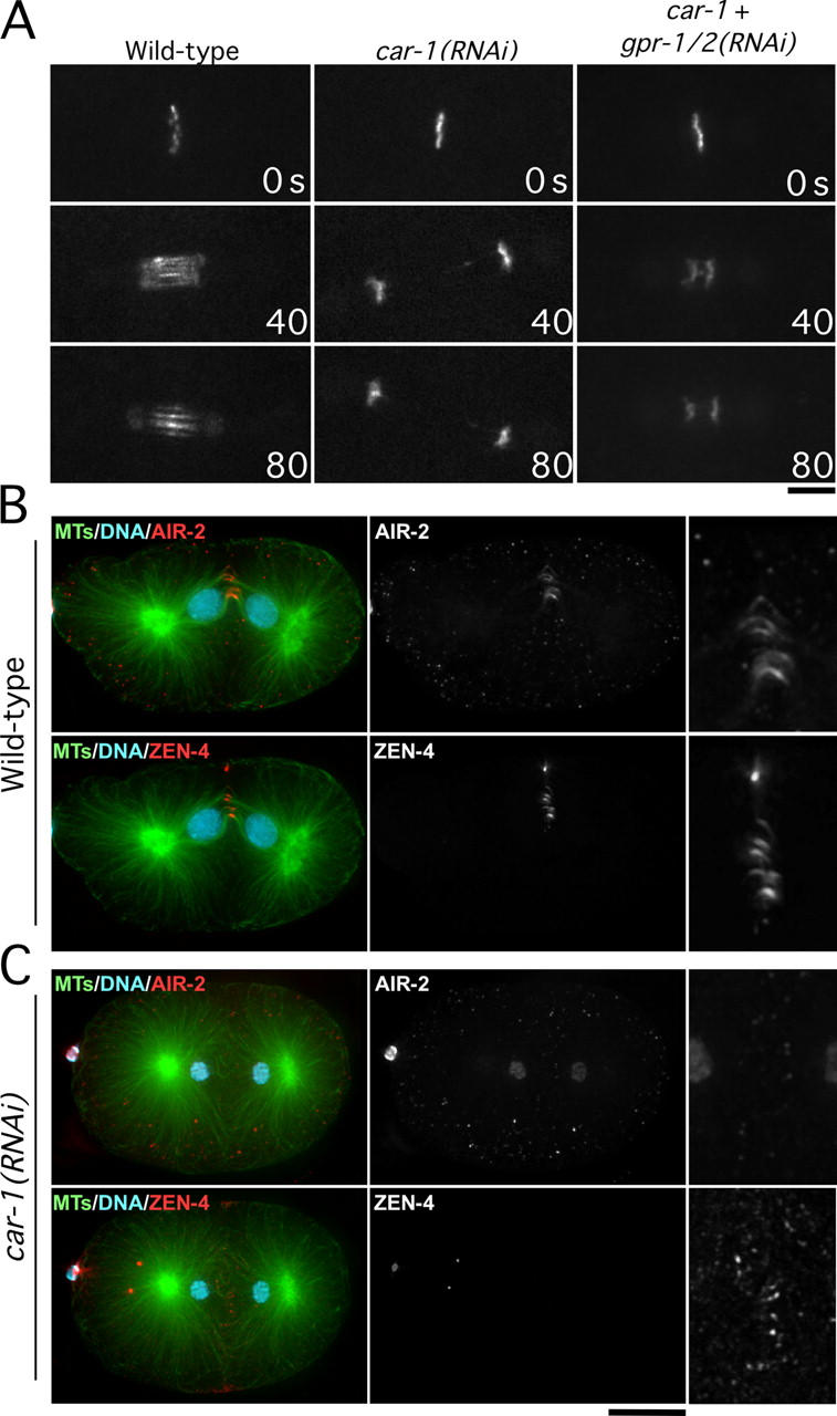
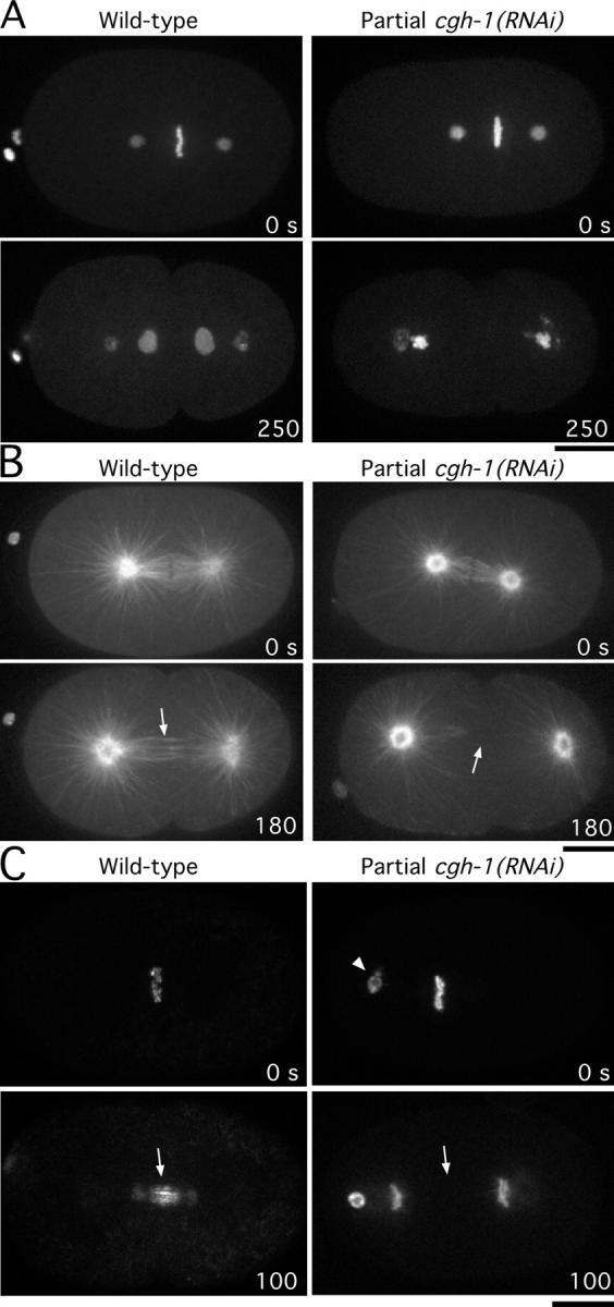
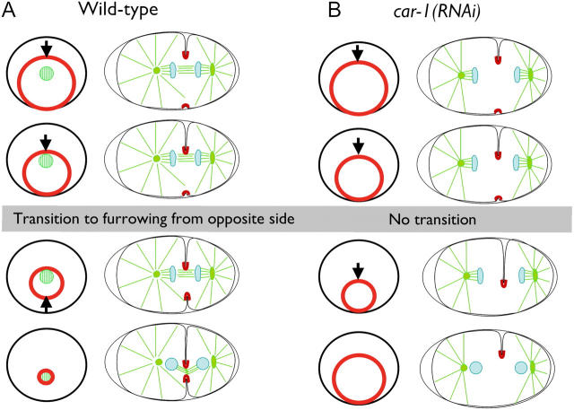
Cytokinesis completes cell division and partitions the contents of one cell to the two daughter cells. Here we characterize CAR-1, a predicted RNA binding protein that is implicated in cytokinesis. CAR-1 localizes to germline-specific RNA-containing particles and copurifies with the essential RNA helicase, CGH-1, in an RNA-dependent fashion. The atypical Sm domain of CAR-1, which directly binds RNA, is dispensable for CAR-1 localization, but is critical for its function. Inhibition of CAR-1 by RNA-mediated depletion or mutation results in a specific defect in embryonic cytokinesis. This cytokinesis failure likely results from an anaphase spindle defect in which interzonal microtubule bundles that recruit Aurora B kinase and the kinesin, ZEN-4, fail to form between the separating chromosomes. Depletion of CGH-1 results in sterility, but partially depleted worms produce embryos that exhibit the CAR-1-depletion phenotype. Cumulatively, our results suggest that CAR-1 functions with CGH-1 to regulate a specific set of maternally loaded RNAs that is required for anaphase spindle structure and cytokinesis.


Figures

Figure 1.
Figure 1.
Cytokinesis fails in car- 1 mutant embryos. (A) Schematic illustration of the domain structure of CAR-1. Polyclonal antibodies were generated against the underlined region. (B) Selected panels from time-lapse DIC sequences of wild-type embryos (left column), car-1(RNAi) embryos (middle column), and car-1(tm1753) mutant embryos (right column). Time in seconds after apparent chromosome alignment is indicated in the lower right corner of each panel. (See also Videos 1–3). Bar, 10 μm.
Figure 2.
Figure 2.
CAR-1 localizes to P-granules and additional smaller cytoplasmic particles. (A) Projected three-dimensional datasets of fixed embryos at the indicated cell cycle stages stained for CAR-1 (left column) and PGL-1 (middle column). Merged images with CAR-1 in green, PGL-1 in red, and DNA in blue also are shown. Panels in the right column are of the boxed area magnified 3× relative to the adjacent images. Arrowheads point to examples of juxtaposed CAR-1– and PGL-1–containing particles during meiosis II. Arrows identify smaller cytoplasmic CAR-1–containing particles that lack PGL-1. Bar, 10 μm. (B) No CAR-1 staining is detected in car-1(RNAi) embryos. Bar, 10 μm.
Figure 3.
Figure 3.
The atypical Sm domain is not required for CAR-1 localization but is essential for its function. (A) Schematic illustrations of the domain structure of two fusion proteins whose functionality was compared. A GFP-containing localization and purification (GFPLAP) tag was fused to full-length CAR-1 (top) or a truncated version of CAR-1 lacking the NH2-terminal Sm domain (bottom). The S-peptide sequence, a component of the GFPLAP tag used for biochemical purification, also is indicated. (B) Projected three-dimensional dataset of a living wild-type embryo at the four-cell stage expressing GFPLAP:CAR-1. Bar, 10 μm (see also Video 4). (C) Single section spinning disc confocal images of living prometaphase embryos expressing GFPLAP:CAR-1 (left) or GFPLAP:CAR-1ΔN (right) are shown after depletion of endogenous CAR-1 using car-1 3′UTR RNAi. Bar, 10 μm. Western blots of extracts prepared from GFPLAP:CAR-1–expressing worms (D) or GFPLAP:CAR-1ΔN–expressing worms (E) that have been depleted specifically of endogenous CAR-1 by RNAi against the car-1 3′UTR. Serial dilutions of extracts prepared from untreated worms expressing GFPLAP:CAR-1 or GFPLAP:CAR-1ΔN were loaded to quantify depletion levels. (F) Wild-type (N2), GFPLAP:CAR-1–expressing, and GFPLAP:CAR-1ΔN–expressing hermaphrodites that were injected with dsRNA targeted against the car-1 3′UTR to deplete the endogenous protein were scored for brood size and embryonic lethality.
Figure 4.
Figure 4.
CAR-1 copurifies with two widely conserved RNA-binding proteins. (A) Silver stained gel of proteins eluted from S-protein agarose after tandem affinity purification of GFPLAP:CAR-1. Three distinct bands (arrowheads) are present. The two bands labeled with asterisks are contaminants (keratins). (B) Table showing the three proteins identified by solution mass spectrometry. The percent sequence coverage, RNAi phenotype, and molecular weight of each protein is shown. (C) Western blots of CAR-1 immunoprecipitates in the presence (+) or absence (−) of RNaseA, probed with the indicated antibodies. (D) Single sections from deconvolved three-dimensional widefield images of fixed gonads stained for CAR-1 (left column) and PGL-1 (middle column). Merged images with DNA staining (blue) are shown in the right column. Bar, 20 μm. (E) Single section spinning disc confocal imaging of gonads (middle section) from living wild-type or CGH-1–depleted hermaphrodites expressing GFPLAP:CAR-1. Bar, 10 μm.
Figure 5.
Figure 5.
Disruption of cleavage furrow ingression in CAR-1–depleted embryos. (A) Selected panels from time-lapse sequences of wild-type (left column) and CAR-1–depleted (right column) embryos expressing GFP:PHPLC1δ1, which localizes to the plasma membrane. Time in seconds after initiation of furrowing is indicated in the lower right corner of each panel. (See also Video 5.) Bar, 10 μm. Arrows highlight the failure of polar body extrusion. Arrowheads point to secondary furrow, which fails to ingress in CAR-1–depleted embryos. (B) Same as in A, except only a 7.3-μm wide vertical section containing the furrow is shown for each time point. Bar, 5 μm. (C) Kymographs of the regions shown in B, comparing furrow ingression in wild-type and CAR-1–depleted embryos (see Methods and materials for details on kymograph construction). The slope of the yellow line indicates the initial rate of primary furrow ingression, which is identical in wild-type and CAR-1–depleted embryos. In wild-type embryos, the primary furrow encounters the midbody and ceases to ingress (time indicated by red dashed line). At a similar time in CAR-1–depleted embryos, the primary furrow slows (cyan line) but does not stop. In wild-type embryos, a secondary furrow begins to ingress from the opposite side of the embryo (pink line) as the primary furrow ceases its inward movement. In CAR-1–depleted embryos, no ingression of a secondary furrow is observed. Bar, 5 μm.
Figure 6.
Figure 6.
CAR-1 depletion results in a pronounced defect in anaphase spindle structure. (A) Selected panels from time-lapse sequences of wild-type (left column) and CAR-1–depleted (right column) embryos expressing GFP:α-tubulin. Time in seconds after chromosome alignment is indicated in the lower right corner of each panel. Arrows indicate the region between the segregated chromosomes where interzonal microtubule bundles normally form (see also Video 7). Bar, 10 μm. (B) Selected panels from time-lapse sequences of wild-type (left column) and CAR-1–depleted (right column) embryos expressing GFP:histone H2B and GFP:γ-tubulin. Time in seconds after chromosome alignment is indicated in the lower right corner of each panel. Arrows point to chromosome bridges that become evident after anaphase onset in CAR-1–depleted embryos. (See also Video 8.) Bar, 10 μm. (C) The distance between spindle poles was tracked for 15 wild-type (WT) and 19 CAR-1–depleted embryos imaged as in A. Average pole-to-pole distance is plotted versus time after NEBD (nuclear envelope breakdown). Error bars represent the SEM with a confidence interval of 0.95. For reference, the time of onset of chromosome segregation is indicated. Embryos in which extrusion of the second polar body failed and extra chromatin was present in the mitotic spindle (n = 6/50) were not used for the analysis of spindle length.
Figure 7.
Figure 7.
AIR-2 and ZEN-4 fail to target to interzonal microtubule bundles in CAR-1–depleted embryos. Simultaneous depletion of GPR-1/2, which inhibits pulling forces on the spindle poles and prevents spindle snapping, does not rescue this defect. (A) Selected images from time-lapse sequences of wild-type, car-1(RNAi), and car-1/gpr-1,2 double RNAi embryos expressing GFP:AIR-2. Only the region of the spindle is shown. Time after chromosome alignment (in seconds) is indicated in the lower right corner of each panel. Bar, 5 μm. Projected three-dimensional datasets of fixed wild-type (B) and CAR-1–depleted (C) embryos stained for DNA, microtubules, AIR-2, and ZEN-4. High magnification panels in the right column are magnified 2.5× relative to the adjacent images. The ZEN-4 high magnification image is 10× overexposed. Bar, 10 μm.
Figure 8.
Figure 8.
Partial depletion of CGH-1 phenocopies depletion of CAR-1. Selected panels from time-lapse sequence of wild-type (left column) and CGH-1 partially depleted (right column) embryos expressing GFP:histone H2B and GFP:γ-tubulin (A), GFP:α-tubulin (B), and GFP:AIR-2 (C). Arrows in B highlight the presence or absence of interzonal microtubules. Arrows in C highlight the presence or absence of AIR-2 on interzonal microtubules; the arrowhead points to polar body chromatin that failed to be extruded properly. Time in seconds after chromosome alignment is indicated in the lower right corner of each panel. Bar, 10 μm.
Figure 9.
Figure 9.
Schematic illustrating the asymmetric furrow ingression in wild-type and car-1(RNA i) embryos. End-on and side views are shown. (A) The contractile ring is positioned asymmetrically within the circumference of the cell equator. In a side view, a primary furrow is observed coming in from one side of the embryo. Contact between the primary furrow and the interzonal microtubule bundles between the separated chromosomes triggers the other side of the contractile ring (the secondary furrow) to pull away from the edge of the embryo, and the contractile ring closes down around the compacted interzonal microtubule bundles (the midbody). (B) In car-1(RNAi) embryos, the primary furrow ingresses, but fails to come into contact with interzonal microtubule bundles. This prevents the structural transition that allows the contractile ring to pull away from the side of the embryo opposite the primary furrow. The furrow fails to close completely and cytokinesis fails.

References

    1. Albertson, D. 1984. Formation of the first cleavage spindle in nematode embryos. Dev. Biol. 101:61–72. - PubMed
    1. Anantharaman, V., and L. Aravind. 2004. Novel conserved domains in proteins with predicted roles in eukaryotic cell-cycle regulation, decapping and RNA stability. BMC Genomics. 16:45. - PMC - PubMed
    1. Blower, M.D., M. Nachury, R. Heald, and K. Weis. 2005. A Rae1-containing ribonucleoprotein complex is required for mitotic spindle assembly. Cell. 121:223–234. - PubMed
    1. Bringmann, H., and A.A. Hyman. 2005. A cytokinesis furrow is positioned by two consecutive signals. Nature. 436:731–734. - PubMed
    1. Cheeks, R.J., J.C. Canman, W.N. Gabriel, N. Meyer, S. Strome, and B. Goldstein. 2004. C. elegans PAR proteins function by mobilizing and stabilizing asymmetrically localized protein complexes. Curr. Biol. 14:851–862. - PubMed

Publication types

MeSH terms