Review
. 2005;5(12):1151-70.
doi: 10.2174/156802605774370856.

Combining cytotoxic and immune-mediated gene therapy to treat brain tumors

Affiliations
Review

Combining cytotoxic and immune-mediated gene therapy to treat brain tumors

James F Curtin et al. Curr Top Med Chem. 2005.

Abstract

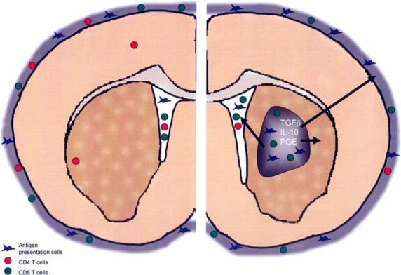
Glioblastoma (GBM) is a type of intracranial brain tumor, for which there is no cure. In spite of advances in surgery, chemotherapy and radiotherapy, patients die within a year of diagnosis. Therefore, there is a critical need to develop novel therapeutic approaches for this disease. Gene therapy, which is the use of genes or other nucleic acids as drugs, is a powerful new treatment strategy which can be developed to treat GBM. Several treatment modalities are amenable for gene therapy implementation, e.g. conditional cytotoxic approaches, targeted delivery of toxins into the tumor mass, immune stimulatory strategies, and these will all be the focus of this review. Both conditional cytotoxicity and targeted toxin mediated tumor death, are aimed at eliminating an established tumor mass and preventing further growth. Tumors employ several defensive strategies that suppress and inhibit anti-tumor immune responses. A better understanding of the mechanisms involved in eliciting anti-tumor immune responses has identified promising targets for immunotherapy. Immunotherapy is designed to aid the immune system to recognize and destroy tumor cells in order to eliminate the tumor burden. Also, immune-therapeutic strategies have the added advantage that an activated immune system has the capability of recognizing tumor cells at distant sites from the primary tumor, therefore targeting metastasis distant from the primary tumor locale. Pre-clinical models and clinical trials have demonstrated that in spite of their location within the central nervous system (CNS), a tissue described as 'immune privileged', brain tumors can be effectively targeted by the activated immune system following various immunotherapeutic strategies. This review will highlight recent advances in brain tumor immunotherapy, with particular emphasis on advances made using gene therapy strategies, as well as reviewing other novel therapies that can be used in combination with immunotherapy. Another important aspect of implementing gene therapy in the clinical arena is to be able to image the targeting of the therapeutics to the tumors, treatment effectiveness and progression of disease. We have therefore reviewed the most exciting non-invasive, in vivo imaging techniques which can be used in combination with gene therapy to monitor therapeutic efficacy over time.


Figures

Fig. (1)
Fig. (1)
Immune cell surveillance of the normal and tumor bearing brain. In the normal brain, only memory CD4+ T-cells are found within the brain parenchyma while other immune cell types are restricted to the cerebrospinal fluid and meningeal layers. Tumors affect the local and systemic immune environment to evade immune detection by producing cytokines like TGFβ, IL-10 and PGE. CD4+ T-cell numbers are reduced systemically. Meanwhile tumors are infiltrated with macrophages and CD8+ T-cells whose normal immunological functions are blocked in the tumor environment.
Fig. (2)
Fig. (2)
Immunotherapy of brain tumors using RAdFlt3L and RAdTK. A. Survival curve from rats treated ten days after tumor implantation with replication deficient adenoviruses. RAdTK+Flt3L results in tumor regression in 80% of animals (modified from Cancer Research [169]). B. Brain section from a glioma survivor 240 days after tumor implantation followed by RAdTK and RAdFlt3L treatment. Brain sections were stained with vimentin to detect any reminant of tumor cells. C and D. Brain sections from animals treated with either saline or RAd TK+Flt3L 10 days after tumor implantation. While 80% of animals treated with RAd TK+Flt3L survive, neuropathological analysis of those who secumb shows immune cell infiltration that is distinct from saline treated controls. As with human tumors, rat CNS-1 gliomas are heavily infiltrated with macrophages (ED1 staining) regardless of treatment modality. B cells (CD45R staining), cytotoxic T cells (CD8 staining), and Natural Killer cells (CD161a staining) are all increased in treated animals compared to controls.
Fig. (3)
Fig. (3)
Targeted toxins for the treatment of glioma. Human brain tumors have been shown to overexpress several receptors, including urokinase-type plasminogen activator receptor, transferring receptor, pleiotropic immunoregulatory cytokine receptors and growth factor receptors. The expression of these receptors seems to be more abundant in malignant tumors, than in benign, slow growing tumors. Since these receptors are virtually absent in the normal brain, they have been targeted in several therapeutic approaches in the treatment of glioma, to avoid toxicity to normal brain tissue. Ligands of these receptors, such as IL-13, IL-4, uPA, transferrin, EGF and TGF have been fused to the catalytic and translocation domains of highly cytotoxic bacterial products, including Pseudomonas and Diphteria toxins (T), in order to kill selectively malignant glioma cells, but preserving surrounding normal brain tissue.
Fig. (4)
Fig. (4)
Stratagies for live animal imaging A. HSV1-Thymidine kinase (TK) is expressed by a first generation adenoviral vector (RAd) and catalyzes the phosphorylation of the prodrug ganciclovir (GCV) to generate GCV-monophosphate (GCV-P). Cellular kinases further phosphorylate to form a nucleotide analogue GCV-triphosphate (GCV-P-P-P) which interferes with DNA synthesis and induces apoptosis in dividing cells. In addition, the HSV1-TK protein interacts with the imaging substrate (124I)-labelled-2’-fluoro-2′-deoxy-1-ß-D-arabinofuranosyl-5-iodouracil (I*-FIAU) to form a phosphorylated version of the molecule (I*-FIAU-P) which can be used for PET imaging. I*-FIAU is membrane permeable and can diffuse freely through the plasma membrane of cells. However, following phosphorylation by TK, I*-FIAU-P becomes hydrophylic and remains trapped in the cell expressing TK. Consequently, the concentration of radioactive 124I increases only in cells expressing TK and is rapidly cleared from the rest of the body. B. A RAd expressing Firefly Luciferase was injected into the striatum of a mouse. Seven days later the mouse was injected with the substrate D-Luciferin and the animal was imaged using a CCD-based in vivo imaging system (IVIS, Xenogen). The intensity of light production was evaluated and is represented using a color scale with red being regions with most intensity and blue being regions with lowest intensity, as indicated on the scale to the right of the image. C. A RAd expressing monomeric red fluorescent protein (mRFP) was infected onto a monolayer of 293 cells. The monolayer was imaged using a inverted fluorescent microscope 24 hours after infection (Axiovert 200, ZEISS).

References

    1. Ali-Osman F. Contemporary Cancer Research. Humana Press; Totowa: 2005. Brain Tumors.
    1. Legler JM, Ries LA, Smith MA, Warren JL, Heineman EF, Kaplan RS, Linet MS. Cancer surveillance series [corrected]: brain and other central nervous system cancers: recent trends in incidence and mortality. J. Natl. Cancer Inst. 1999;91(16):1382–90. - PubMed
    1. Surawicz TS, Davis F, Freels S, Laws ER, Jr., Menck HR. Brain tumor survival: results from the National Cancer Data Base. J. Neurooncol. 1998;40(2):151–60. - PubMed
    1. Platten M, Wick W, Weller M. Malignant glioma biology: role for TGF-beta in growth, motility, angiogenesis, and immune escape. Microsc. Res. Tech. 2001;52(4):401–10. - PubMed
    1. Yang L, Ng KY, Lillehei KO. Cell-mediated immunotherapy: a new approach to the treatment of malignant glioma. Cancer Control. 2003;10(2):138–47. - PubMed

Publication types

MeSH terms