

Botulinum toxin treatment of extraocular muscles in rabbits results in increased myofiber remodeling
- PMID: 16249488
- PMCID: PMC1847582
- DOI: 10.1167/iovs.05-0549
Botulinum toxin treatment of extraocular muscles in rabbits results in increased myofiber remodeling
Abstract
Purpose: Botulinum toxin A (Botox) is commonly used for strabismus treatment. Although other muscles atrophy after intramuscular injection with Botox, extraocular muscles (EOMs) do not. A continuous process of myonuclear addition in normal uninjured adult myofibers in rabbit EOMs was studied. In this study, the effect of Botox-induced muscle paralysis on myofiber remodeling in adult EOMs was examined.
Methods: The superior rectus muscles of adult rabbits were each injected with 5 units of Botox. The contralateral muscle received injections of saline only. Bromodeoxyuridine (BrdU) was administered for various periods after Botox treatment, followed by various BrdU-free periods. Myonuclear addition, the number of BrdU-positive satellite cells, and the number of MyoD-positive satellite cells were quantified, as were alterations in expression of immature myosins.
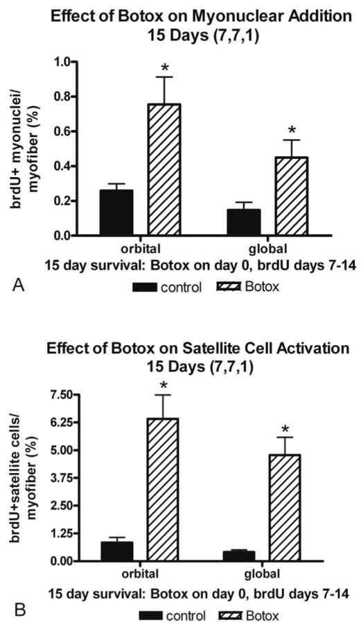
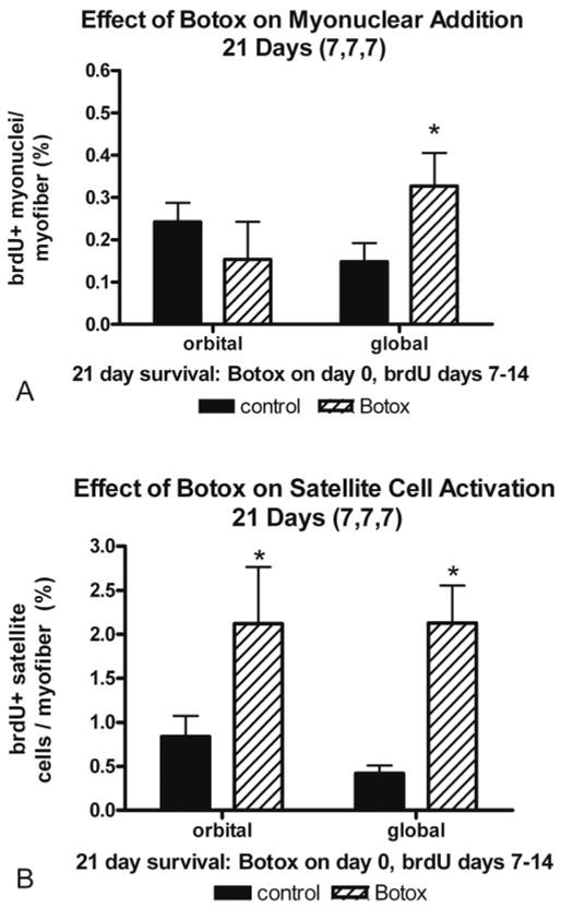
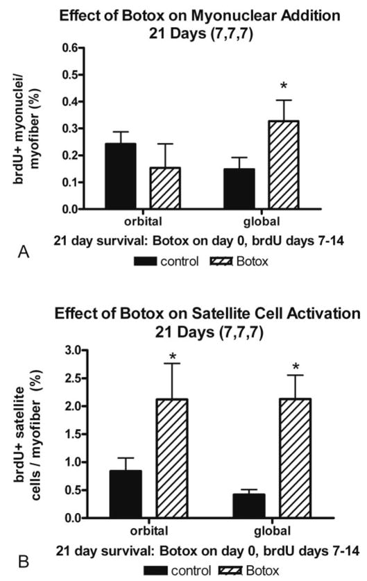
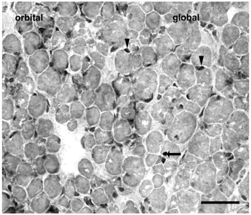
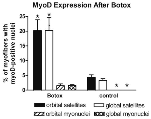
Results: Intramuscular injection of Botox resulted in a significant increase in both the number of BrdU-positive myonuclei and satellite cells. MyoD expression in both satellite cells and myonuclei was significantly increased after Botox injection in EOMs. In Botox-treated EOMs, an increased number of myofibers positive for the neonatal myosin heavy chain (MyHC) isoform was detected in the orbital layer.
Conclusions: Botox-induced EOM paralysis resulted in a significant short-term increase in satellite cell activation and myonuclear addition in single myofibers in adult rabbit EOMs compared with control muscles. The appearance of MyoD-positive myonuclei suggests that protein synthesis becomes upregulated after Botox injection, and this, in turn, may help explain the minimal effects on myofiber size in EOMs after Botox injection. Understanding the effect of Botox on satellite cell activation and myonuclear addition in existing myofibers may suggest new ways to maximize the clinical effectiveness of Botox in patients with strabismus.
Figures
References
-
- Scott AB, Kennedy RA, Stubbs HA. Botulinum toxin injection as a treatment for blepharospasm. Arch Ophthalmol. 1985;103:347–350. - PubMed
-
- Brin MF, Fahn S, Moskowitz D, et al. Localized injections of Botulinum toxin for treatment of focal dystonia and hemifacial spasm. Mov Disord. 1987;2:237–254. - PubMed
-
- Koman LA, Mooney JF, Smith B, Goodman A, Mulvaney T. Management of cerebral palsy with botulinum-A toxin: preliminary investigation. J Pediatr Orthop. 1993;13:489–495. - PubMed
-
- Scott AB. Botulinum toxin injection into extraocular muscles as an alternative to strabismus surgery. Ophthalmology. 1980;87:1044–1049. - PubMed
-
- Scott AB, Rosenbaum A, Collins CC. Pharmacologic weakening of extraocular muscles. Invest Ophthalmol. 1973;12:924–927. - PubMed
Publication types
MeSH terms
Substances
Grants and funding
LinkOut - more resources
Full Text Sources
Medical
