

Axial stent strut angle influences wall shear stress after stent implantation: analysis using 3D computational fluid dynamics models of stent foreshortening
- PMID: 16250918
- PMCID: PMC1276824
- DOI: 10.1186/1475-925X-4-59
Axial stent strut angle influences wall shear stress after stent implantation: analysis using 3D computational fluid dynamics models of stent foreshortening
Abstract
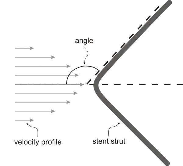
Introduction: The success of vascular stents in the restoration of blood flow is limited by restenosis. Recent data generated from computational fluid dynamics (CFD) models suggest that the vascular geometry created by an implanted stent causes local alterations in wall shear stress (WSS) that are associated with neointimal hyperplasia (NH). Foreshortening is a potential limitation of stent design that may affect stent performance and the rate of restenosis. The angle created between axially aligned stent struts and the principal direction of blood flow varies with the degree to which the stent foreshortens after implantation.
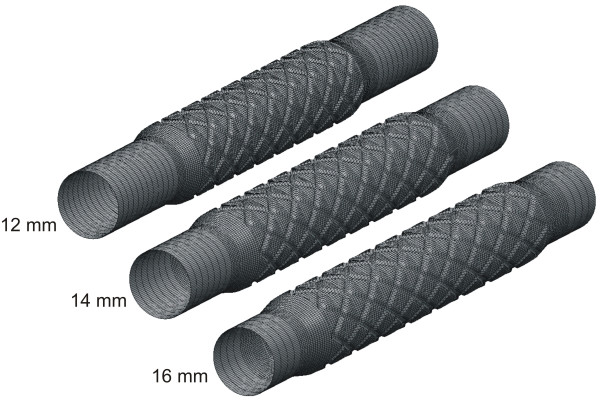
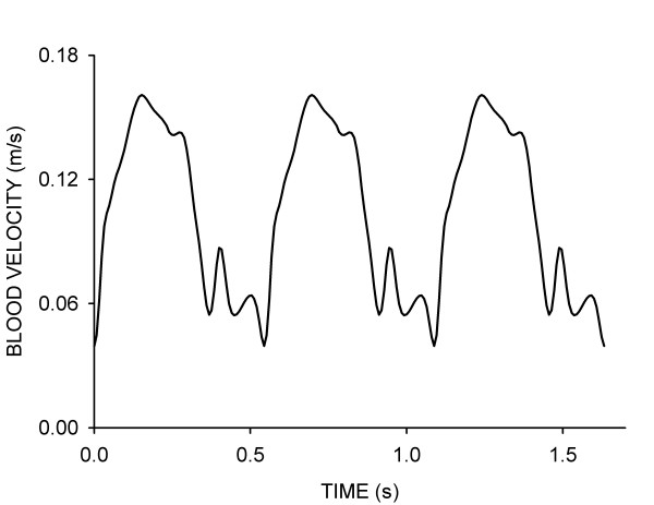
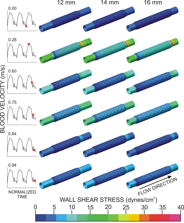
Methods: In the current investigation, we tested the hypothesis that stent foreshortening adversely influences the distribution of WSS and WSS gradients using time-dependent 3D CFD simulations of normal arteries based on canine coronary artery measurements of diameter and blood flow. WSS and WSS gradients were calculated using conventional techniques in ideal (16 mm) and progressively foreshortened (14 and 12 mm) stented computational vessels.
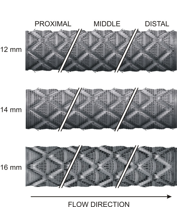
Results: Stent foreshortening increased the intrastrut area of the luminal surface exposed to low WSS and elevated spatial WSS gradients. Progressive degrees of stent foreshortening were also associated with strut misalignment relative to the direction of blood flow as indicated by analysis of near-wall velocity vectors.
Conclusion: The current results suggest that foreshortening may predispose the stented vessel to a higher risk of neointimal hyperplasia.
Figures
Similar articles
-
Alterations in regional vascular geometry produced by theoretical stent implantation influence distributions of wall shear stress: analysis of a curved coronary artery using 3D computational fluid dynamics modeling.Biomed Eng Online. 2006 Jun 16;5:40. doi: 10.1186/1475-925X-5-40. Biomed Eng Online. 2006. PMID: 16780592 Free PMC article.
-
Circumferential vascular deformation after stent implantation alters wall shear stress evaluated with time-dependent 3D computational fluid dynamics models.J Appl Physiol (1985). 2005 Mar;98(3):947-57. doi: 10.1152/japplphysiol.00872.2004. Epub 2004 Nov 5. J Appl Physiol (1985). 2005. PMID: 15531564
-
Blood flow in stented arteries: a parametric comparison of strut design patterns in three dimensions.J Biomech Eng. 2005 Aug;127(4):637-47. doi: 10.1115/1.1934122. J Biomech Eng. 2005. PMID: 16121534
-
[Survey of coronary stents development for restenosis prevention].Zhongguo Yi Liao Qi Xie Za Zhi. 2009 Nov;33(6):429-34. Zhongguo Yi Liao Qi Xie Za Zhi. 2009. PMID: 20352916 Review. Chinese.
-
Computational fluid dynamics and stent design.Artif Organs. 2002 Jul;26(7):614-21. doi: 10.1046/j.1525-1594.2002.07084.x. Artif Organs. 2002. PMID: 12081520 Review.
Cited by
-
Alterations in regional vascular geometry produced by theoretical stent implantation influence distributions of wall shear stress: analysis of a curved coronary artery using 3D computational fluid dynamics modeling.Biomed Eng Online. 2006 Jun 16;5:40. doi: 10.1186/1475-925X-5-40. Biomed Eng Online. 2006. PMID: 16780592 Free PMC article.
-
Effects of stent sizing on endothelial and vessel wall stress: potential mechanisms for in-stent restenosis.J Appl Physiol (1985). 2009 May;106(5):1686-91. doi: 10.1152/japplphysiol.91519.2008. Epub 2009 Mar 19. J Appl Physiol (1985). 2009. PMID: 19299567 Free PMC article.
-
Wall shear stress in intracranial self-expanding stents studied using ultra-high-resolution 3D reconstructions.AJNR Am J Neuroradiol. 2009 Mar;30(3):479-86. doi: 10.3174/ajnr.A1396. Epub 2008 Nov 27. AJNR Am J Neuroradiol. 2009. PMID: 19039050 Free PMC article.
-
Propensity-matched analysis of long-term clinical results after ostial circumflex revascularisation.Heart. 2023 Aug 11;109(17):1302-1309. doi: 10.1136/heartjnl-2022-322204. Heart. 2023. PMID: 37217296 Free PMC article.
-
Compound ex vivo and in silico method for hemodynamic analysis of stented arteries.PLoS One. 2013;8(3):e58147. doi: 10.1371/journal.pone.0058147. Epub 2013 Mar 13. PLoS One. 2013. PMID: 23516442 Free PMC article.
References
-
- Farb A, Weber DK, Kolodgie FD, Burke AP, Virmani R. Morphological predictors of restenosis after coronary stenting in humans. Circulation. 2002;105:2974–2980. doi: 10.1161/01.CIR.0000019071.72887.BD. - DOI - PubMed
-
- Duda SH, Wiskirchen J, Tepe G, Bitzer M, Kaulich TW, Stoeckel D, Claussen CD. Physical properties of endovascular stents: an experimental comparison. Journal of Vascular and Interventional Radiology. 2000;11:645–654. - PubMed
-
- Kalmar G, Hubner F, Voelker W, Hutzenlaub J, Teubner J, Poerner T, Suselbeck T, Borggrefe M, Haase KK. Radial force and wall apposition of balloon-expandable vascular stents in eccentric stenoses: an in vitro evaluation in a curved vessel model. Journal of Vascular and Interventional Radiology. 2002;13:499–508. - PubMed
Publication types
MeSH terms
Grants and funding
LinkOut - more resources
Full Text Sources
Other Literature Sources
Research Materials
Miscellaneous
