

Changes in circulating mesenchymal stem cells, stem cell homing factor, and vascular growth factors in patients with acute ST elevation myocardial infarction treated with primary percutaneous coronary intervention
- PMID: 16251230
- PMCID: PMC1860647
- DOI: 10.1136/hrt.2005.069799
Changes in circulating mesenchymal stem cells, stem cell homing factor, and vascular growth factors in patients with acute ST elevation myocardial infarction treated with primary percutaneous coronary intervention
Abstract
Objective: To investigate the spontaneous occurrence of circulating mesenchymal stem cells (MSC) and angiogenic factors in patients with ST elevation acute myocardial infarction (STEMI) treated with primary percutaneous coronary intervention (PCI).
Design: In 20 patients with STEMI, blood samples were obtained on days 1, 3, 7, 14, 21, and 28 after the acute PCI. Fifteen patients with a normal coronary angiography formed a control group. MSC (CD45-/CD34-), plasma stromal derived factor 1 (SDF-1), vascular endothelial growth factor A (VEGF-A), and fibroblast growth factor 2 (FGF-2) were measured by multiparametric flow cytometry and enzyme linked immunosorbent assay (ELISA).
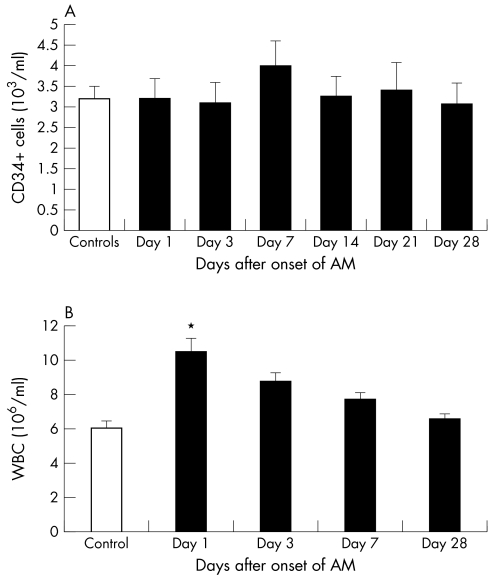
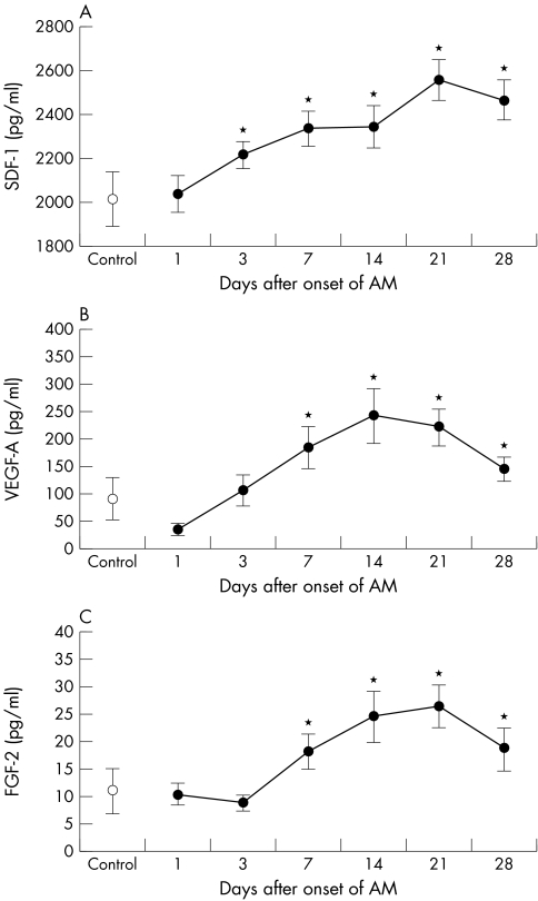
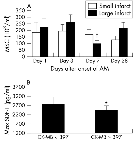
Results: Circulating CD45-/CD34- cells were significantly decreased on day 7 compared with day 3. Cell counts normalised one month after the acute onset of STEMI. The changes were mainly seen in patients with a large infarction. Plasma SDF-1 increased significantly from day 3 to day 28, and VEGF-A and FGF-2 increased significantly from day 7 to day 28.
Conclusions: Spontaneous sequential fluctuations in MSC and the increase in vascular growth factor concentrations after STEMI suggest that the optimal time for additional stem cell therapy is three weeks after a myocardial infarction to obtain the maximum effects by stimulating endogenous growth factors on the delivered stem cells.
Conflict of interest statement
Conflict of interest: Nothing declared.


References
-
- Orlic D, Kajstura J, Chimenti S.et al Bone marrow cells regenerate infracted myocardium. Nature 2001410701–705. - PubMed
-
- Asahara T, Masuda H, Takahashi T.et al Bone marrow origin of endothelial progenitor cells responsible for postnatal vasculogenesis in physiological and pathological neovascularization. Circ Res 199985221–228. - PubMed
-
- Schachinger V, Assmus B, Britten M B.et al Transplantation of progenitor cells and regeneration enhancement in acute myocardial infarction: final one‐year results of the TOPCARE‐AMI trial. J Am Coll Cardiol 2004441690–1699. - PubMed
-
- Jørgensen E, Ripa R S, Wang Y.et al Instent Neo‐intimal hyperplasia after stem cell mobilization by granulocyte‐colony stimulating factor: preliminary intracoronary ultrasound results from a double‐blind randomized placebo‐controlled study of patients treated with percutaneous coronary intervention for ST‐elevation myocardial infarction (STEMMI trial). Int J Cardiol. (in press) - PubMed
Publication types
MeSH terms
Substances
LinkOut - more resources
Full Text Sources
Other Literature Sources
Medical
Research Materials
Miscellaneous