. 2005 Nov 8;102(45):16275-80.
doi: 10.1073/pnas.0508105102. Epub 2005 Oct 26.

Regulation of the Caenorhabditis elegans oxidative stress defense protein SKN-1 by glycogen synthase kinase-3

Affiliations

Regulation of the Caenorhabditis elegans oxidative stress defense protein SKN-1 by glycogen synthase kinase-3

Jae Hyung An et al. Proc Natl Acad Sci U S A. .

Abstract

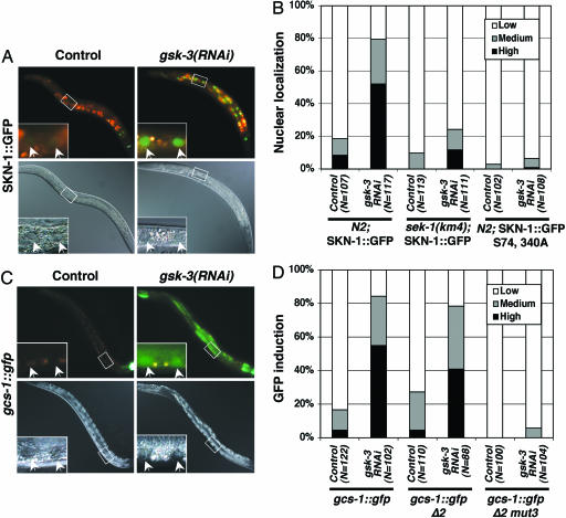
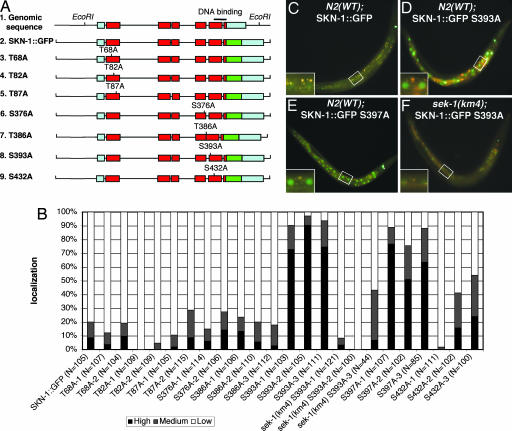
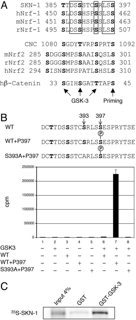
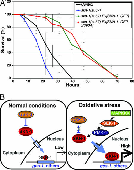
Oxidative stress plays a central role in many human diseases and in aging. In Caenorhabditis elegans the SKN-1 protein induces phase II detoxification gene transcription, a conserved oxidative stress response, and is required for oxidative stress resistance and longevity. Oxidative stress induces SKN-1 to accumulate in intestinal nuclei, depending on p38 mitogen-activated protein kinase signaling. Here we show that, in the absence of stress, phosphorylation by glycogen synthase kinase-3 (GSK-3) prevents SKN-1 from accumulating in nuclei and functioning constitutively in the intestine. GSK-3 sites are conserved in mammalian SKN-1 orthologs, indicating that this level of regulation may be conserved. If inhibition by GSK-3 is blocked, background levels of p38 signaling are still required for SKN-1 function. WT and constitutively nuclear SKN-1 comparably rescue the skn-1 oxidative stress sensitivity, suggesting that an inducible phase II response may provide optimal stress protection. We conclude that (i) GSK-3 inhibits SKN-1 activity in the intestine, (ii) the phase II response integrates multiple regulatory signals, and (iii), by inhibiting this response, GSK-3 may influence redox conditions.


Figures

Fig. 1.
Fig. 1.
gsk-3 inhibits SKN-1 localization and SKN-1 target gene expression. (A and B) SKN-1 is localized to intestinal nuclei in gsk-3(RNAi) animals in the absence of stress. N2 Is007[SKN-1::GFP], sek-1(km4) Is007[SKN-1::GFP], and N2 Ex[SKN-1::GFP S74, 340A] L4 animals were placed onto nematode growth medium plates containing E. coli HT115 that carried either gsk-3 dsRNA or control (L4440) feeding vectors. The animals were then incubated for 24 h at 20°C and allowed to lay eggs. Their surviving progeny were scored as L4 larvae and young adults for accumulation of SKN-1::GFP in intestinal nuclei as described in ref. . “High” indicates that a strong SKN-1::GFP signal was present in all intestinal nuclei, as in the gsk-3(RNAi) animal shown. “Medium” refers to animals in which nuclear SKN-1::GFP was present at high levels anteriorly or anteriorly and posteriorly but was barely detectable midway through the intestine. sek-1 encodes a C. elegans p38 MAPK kinase and is required for SKN-1 function in the intestine (see Results and Discussion) (28). (C and D) gcs-1::gfp is expressed constitutively in gsk-3(RNAi) animals. gsk-3 RNAi was performed in gcs-1::gfp, gcs-1::gfp Δ2, and gcs-1::gfp Δ2-mut3 strains as in A and B, then intestinal gcs-1::gfp expression was scored in F1 L4 larvae and young adults (11). “High” indicates that gcs-1::gfp was present at high levels anteriorly and was detectable throughout most of the intestine, as in the gsk-3(RNAi) image shown. “Medium” refers to animals in which gcs-1::gfp was present at high levels anteriorly and possibly posteriorly but was not detected in between. The gcs-1::gfp fluorescence apparent in the control image derives from SKN-1-independent pharyngeal expression (11). In A and C, upper images show fluorescence, lower images show Nomarski imaging, and pairs of intestinal nuclei (arrows) are shown in each inset.
Fig. 2.
Fig. 2.
A predicted GSK-3 phosphorylation site prevents constitutive accumulation of SKN-1::GFP in intestinal nuclei. (A) The SKN-1 coding region and mutant transgenic constructs. Predicted coding regions are indicated by red boxes, untranslated regions are indicated by blue boxes, and GFP is indicated by a green box. Potential GSK-3 sites in SKN-1 were predicted by the scansite program (Table 1) (32). These predicted phosphorylated residues and the priming site Ser-397 (see Results and Discussion) were individually substituted with alanine within SKN-1::GFP. (BE) Analysis of transgenic animals. (B) Individual transgenic lines were generated by injecting the transgenes diagrammed in A along with the marker rol-6 (pRF4). These extrachromosomal lines were scored for presence of SKN-1 in intestinal nuclei, as in Fig. 1B. Nuclear SKN-1::GFP was dramatically increased in SKN-1::GFP S393A lines in the WT but not sek-1(km4) background and in SKN-1::GFP S397A lines. (CF) Fluorescent images of representative transgenic animals are shown, as in Fig. 1 A.
Fig. 3.
Fig. 3.
Phosphorylation of SKN-1 by GSK-3. (A) A GSK-3 phosphorylation motif in C. elegans SKN-1 is conserved in mammalian Nrf1 proteins. This motif, which has three predicted GSK-3 sites, is similar in structure to the GSK-3 target motif in β-catenin, in which priming phosphorylation at Ser-45 by casein kinase-1α is followed by sequential GSK-3 phosphorylation at Thr-41, Ser-37, and Ser-33 (26). Predicted compound GSK-3 phosphorylation motifs are also present in mammalian Nrf2 and the orthologous Drosophila protein CNC. Conserved residues in each group are boxed. h, human; m, mouse; r, rat. (B) Phosphorylation of SKN-1 Ser-393 by GSK-3β in vitro. GSK-3β phosphorylates the SKN-1 peptide shown provided that phosphorylated serine is present at position 397. This phosphorylation depends on the presence of Ser-393. cpm incorporated in a representative phosphorylation assay are graphed. (C) SKN-1 binds specifically to a C. elegans GST–GSK-3 fusion protein in vitro.
Fig. 4.
Fig. 4.
SKN-1 protects against oxidative stress. (A) SKN-1::GFP overexpression confers oxidative stress resistance. Transgenically expressed WT and S393A forms of SKN-1::GFP comparably rescue the oxidative stress sensitivity of skn-1(zu67) mutants and provide additional protection against oxidative stress. All animals analyzed carry the rol-6 transgenic marker and were derived from worms that are heterozygous for the DnT1 balancer. Control thus refers to N2;rol-6 progeny that were derived from N2;rol-6;DnT1/+ animals. Individual worms were scored for survival at the times shown after they had been placed on nematode growth medium plates containing 6.2 mM t-butyl hydroperoxide. Under these stress conditions, SKN-1::GFP accumulated in intestinal nuclei and gcs-1::gfp expression was increased (data not shown). This representative experiment involved 40 worms in each group. Error bars indicate standard deviations. P values for skn-1(zu67), skn-1(zu67) Ex[SKN-1::GFP], and skn-1(zu67) Ex[SKN-1::GFP S393A] compared with control were 0.034, 0.007, and 0.009, respectively. (B) A model for regulation of SKN-1 in the intestine. Under normal conditions, SKN-1 is phosphorylated constitutively by GSK-3 and prevented from accumulating in the nucleus. This inhibition depends on the prior priming phosphorylation of SKN-1, which represents an additional negative signal (see Results and Discussion). SKN-1 target genes, represented by gcs-1, are then expressed at very low levels. Under oxidative stress conditions, p38 pathway signaling and PMK-1 phosphorylation of SKN-1 are dramatically increased (28). This signal counteracts inhibition of SKN-1 by GSK-3 and is independently required for SKN-1 to accumulate in nuclei at high levels and induce phase II gene expression. These phosphorylation events are arbitrarily shown as occurring in the cytoplasm. MAPKKK, MAPK kinase kinase.

Similar articles

Cited by

References

    1. Brownlee, M. (2001) Nature 414, 813-820. - PubMed
    1. Sheetz, M. J. & King, G. L. (2002) J. Am. Med. Assoc. 288, 2579-2588. - PubMed
    1. Droge, W. (2002) Physiol. Rev. 82, 47-95. - PubMed
    1. Andersen, J. K. (2004) Nat. Med. 10, S18-S25. - PubMed
    1. Balaban, R. S., Nemoto, S. & Finkel, T. (2005) Cell 120, 483-495. - PubMed

Publication types

MeSH terms

Substances

LinkOut - more resources