. 2005 Dec 6;102(49):17551-8.
doi: 10.1073/pnas.0507856102. Epub 2005 Oct 26.

Regulation of RNA splicing by the methylation-dependent transcriptional repressor methyl-CpG binding protein 2

Affiliations

Regulation of RNA splicing by the methylation-dependent transcriptional repressor methyl-CpG binding protein 2

Juan I Young et al. Proc Natl Acad Sci U S A. .

Erratum in

  • Proc Natl Acad Sci U S A. 2006 Jan 31;103(5):1656

Abstract

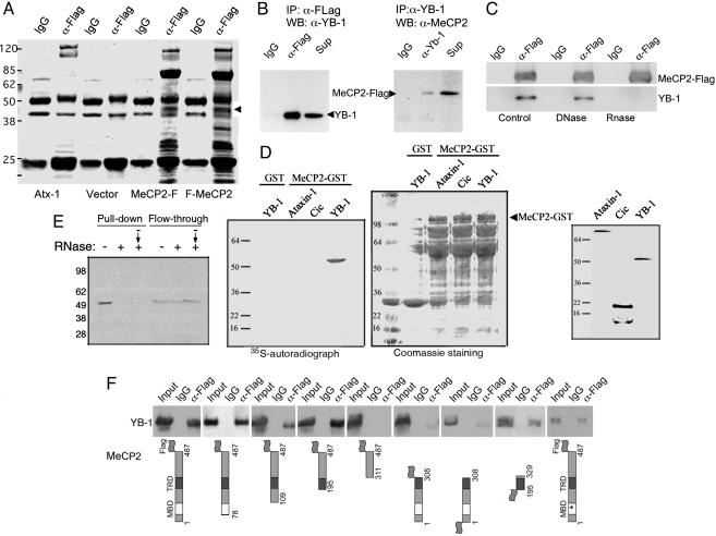
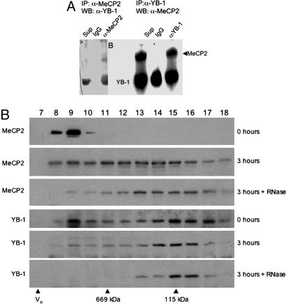
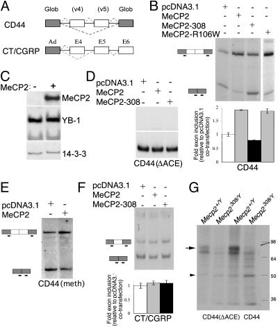
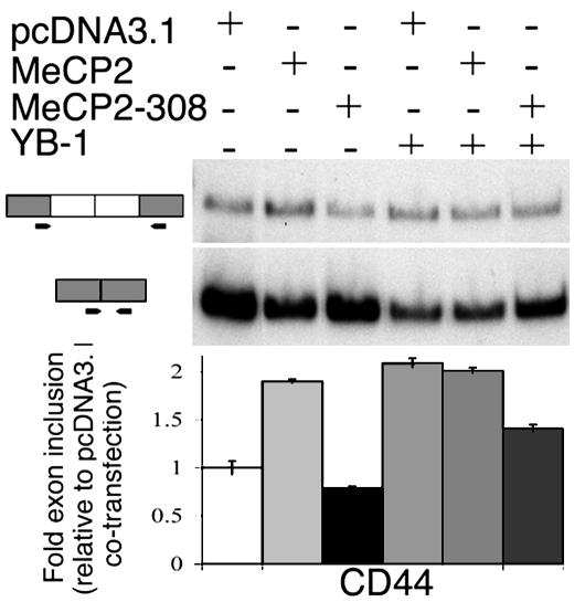
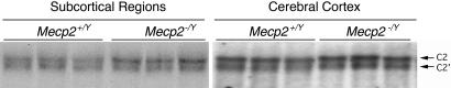
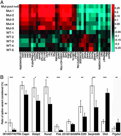
Rett syndrome (RTT) is a postnatal neurodevelopmental disorder characterized by the loss of acquired motor and language skills, autistic features, and unusual stereotyped movements. RTT is caused by mutations in the X-linked gene encoding methyl-CpG binding protein 2 (MeCP2). Mutations in MECP2 cause a variety of neurodevelopmental disorders including X-linked mental retardation, psychiatric disorders, and some cases of autism. Although MeCP2 was identified as a methylation-dependent transcriptional repressor, transcriptional profiling of RNAs from mice lacking MeCP2 did not reveal significant gene expression changes, suggesting that MeCP2 does not simply function as a global repressor. Changes in expression of a few genes have been observed, but these alterations do not explain the full spectrum of Rett-like phenotypes, raising the possibility that additional MeCP2 functions play a role in pathogenesis. In this study, we show that MeCP2 interacts with the RNA-binding protein Y box-binding protein 1 and regulates splicing of reporter minigenes. Importantly, we found aberrant alternative splicing patterns in a mouse model of RTT. Thus, we uncovered a previously uncharacterized function of MeCP2 that involves regulation of splicing, in addition to its role as a transcriptional repressor.


Figures

Fig. 1.
Fig. 1.
YB-1 is a MeCP2-associated protein. (A) Coomassie blue staining of anti-FLAG immunoprecipitates from Neuro2A cells transiently transfected with C- or N-terminally FLAG-tagged MeCP2, MeCP2-F, and F-MeCP2, respectively. The arrowhead indicates the 50-kDa band corresponding to YB-1 that is not immunoprecipitated from cells transfected with empty vector or with a plasmid expressing an unrelated nuclear protein (Atx-1, ataxin-1). (B) FLAG-tagged MeCP2 was transfected into Neuro2A cells, immunoprecipitated with unrelated (IgG, anti-HA), anti-FLAG (Left), or anti-YB-1 (Right) antibodies. The immunoprecipitates were analyzed by Western blot for YB-1 (Left) or MeCP2 (Right). Sup, supernatant. (C) Western blot analysis for the identification of MeCP2-FLAG or YB-1, in anti-FLAG immunoprecipitates from Neuro2A cells transfected with MeCP2-FLAG, with or without DNase or RNase treatment of the extracts. RNase treatment disrupts the MeCP2–YB-1 complex. (D) MeCP2-GST was incubated with IVTT, YB-1, or the unrelated proteins ataxin-1 and capicua. MeCP2 interacts specifically only with YB-1, despite comparable protein loading, visualized by Coomassie blue staining (GST-MeCP2 runs at ≈100 kDa) and comparable input of IVTT products (Right). (E) Pretreatment of the IVTT YB-1 with RNase before incubation with MeCP2-GST (+) and treatment of the incubated mixture, including MeCP2-GST and IVTT YB-1, (– → +) prevents YB-1's pull down by MeCP2-GST. (F) The region of MeCP2 that mediates the interaction with YB-1 was identified by anti-FLAG immunoprecipitation from Neuro2A cells transfected with FLAG-tagged MeCP2 deletion constructs.
Fig. 2.
Fig. 2.
MeCP2 and YB-1 interact in vivo in an RNA-dependent manner. (A) Endogenous co-IP of YB-1 and MeCP2 from CEM-CCRF cells. Immunoprecipitation with an unrelated antibody (IgG, anti-HA), anti-N terminus MeCP2 antibody (Left), or anti-YB-1 (Right) antibodies was followed by Western blotting with anti-YB1 (Left) or anti-C terminus MeCP2 (Right) antibody. (B) Western blot analysis for MeCP2 and YB-1 of fractions 7–18 from Superose 6 size exclusion chromatography of mouse brain nuclear extracts. Fraction numbers are indicated on top; void volume and standards are indicated at the bottom. Extracts were subjected to chromatography immediately upon thawing (0 h) or after incubation for 3 h on ice either with (3 h + RNase) or without (3 h) the addition of RNases A and T1, indicated on the right. Endogenous RNase activity in the 3-h incubation likely resulted in some RNA degradation and, thus, a shift in MeCP2 and YB-1 compared with extracts loaded immediately (0 h). This shift is much more dramatic and tight when exogenous RNase is added to the extracts.
Fig. 3.
Fig. 3.
MeCP2 regulates splicing of alternative exons. (A) Schematic of the splicing reporter minigenes. (B) HeLa cells were cotransfected with a CD44-derived splicing reporter, together with vector (pcDNA3.1), MeCP2, MeCP2-308, or MeCP2-R106W plasmids. Radiolabeled RT-PCR was performed to detect spliced transcripts. (Upper) Representative RT-PCR autoradiographs. (Lower) Mean (± SD, n ≥ 3) quantifications of the fold exon inclusion, relative to the control DNA. (C) Subcellular fractionation, followed by Western analysis of MeCP2 and endogenous YB-1, indicates that nuclear levels of YB-1 are not altered in MeCP2-transfected cells. (D) HeLa cells were cotransfected with a CD44 minigene deleted for the YB-1 binding element (ΔACE), along with YB-1 and pcDNA3.1, MeCP2, or MeCP2-308. Deletion of the AC-rich element turned the minigene insensitive to cotransfection of wild-type or mutant MeCP2. (E) HeLa cells were cotransfected with an in vitro methylated CD44-derived splicing reporter, together with a vector (pcDNA3.1) or MeCP2. Shown are RT-PCR autoradiographs. In vitro methylation of the CD44 splicing reporter resulted in transcriptional repression by MeCP2, therefore, the amount of the CD44 splicing reporter used in this particular experiments was 10 times the amount used in experiments shown in B and D. The MeCP2-dependent increase in exon inclusion was quantitatively similar to that of the unmethylated CD44 splicing reporter. (F) HeLa cells were cotransfected with a CT/CGRP-derived minigene, along with pcDNA3.1, MeCP2, or MeCP2-308. Splicing of this minigene was not modified by overexpression of MeCP2. (G) 32P-labeled CD44 or CD44(ΔACE) mRNA were incubated with nuclear brain extracts prepared from wild-type or Mecp2308/Y mutant mice, UV irradiated, fractionated by SDS/PAGE, and exposed to x-ray film. Both CD44 mRNAs, wild type and mutant, cross linked to two bands of ≈50 (arrowhead) and 80 (arrow) kDa. The labeled 80-kDa band is absent from the mutant extracts. In addition, the mutation of the ACE element in CD44 decreases its affinity for YB-1 and MeCP2.
Fig. 4.
Fig. 4.
HeLa cells cotransfected a CD44 minigene, along with YB-1 and pcDNA3.1, MeCP2, or MeCP2-308. Mutant MeCP2 (MeCP2-308) antagonizes YB-1 exon-inclusion promoting activity. (Upper) Representative RT-PCR autoradiographs. (Lower) Mean (±SD, n ≥ 3) quantifications of the fold exon inclusion, relative to the plasmid control DNA cotransfection.
Fig. 5.
Fig. 5.
MeCP2 regulates NR1 mRNA splicing. Total RNA from brain regions derived from wild-type or Mecp2–[supi]/Y mice was subjected to Northern analysis. Hybridization with an NR1 probe detects both the C2 and C2′ containing mRNA variants (arrows) and shows different ratios of the two variants in Mecp2–/Y vs. wild type (Mecp2+/Y) specifically in the subcortical samples. Note that both isoforms are equally abundant in wild-type samples, whereas the C2 isoform is more abundant in the mutant samples. No differences were observed in cortex-derived mRNA.
Fig. 6.
Fig. 6.
Aberrant splicing in a mouse model of RTT. (A) Supervised clustering of individual mouse samples based on splice monitoring. For every mutually exclusive splicing event identified, we designed a pair of probes to monitor the inclusion and the exclusion of the event. As an example, a cassette exon would have one probe to monitor the exon and one probe to monitor the junction between adjacent exons. To isolate splicing changes beyond gene expression changes, relative gene expression, as monitored by separate probes, was subtracted from each probe. Plotted is the log10-ratio-to-pool difference between probe pairs. Splicing events were selected that distinguish mutant from wild-type (wt) mouse brain samples with a t test (P < 0.0005). The CD44 variants (second to fifth from left) can distinguish samples. (B) Relative quantification of splice variant-specific mRNA by real-time PCR. The relative abundance of a set of splice variants found to be differentially expressed by microarray analysis were analyzed in cerebral cortices of six mice of wild-type (open bars) or mutant (filled bars) genotypes. Depicted are means of triplicate experiments for six mice of each genotype (±SD). *, P < 0.05; **, P < 0.03; ***, P < 0.01.

Similar articles

Cited by

References

    1. Hagberg, B., Aicardi, J., Dias, K. & Ramos, O. (1983) Ann. Neurol., 14, 471–479. - PubMed
    1. Rett, A. (1966) Wien. Med. Wochenschr., 116, 723–726. - PubMed
    1. Einspieler, C., Kerr, A. M. & Prechtl, H. F. (2004) Pediatr. Res., 57, 696–700. - PubMed
    1. Neul, J. L. & Zoghbi, H. Y. (2004) Neuroscientist 10, 118–128. - PubMed
    1. Weaving, L. S., Ellaway, C. J., Gecz, J. & Christodoulou, J. (2005) J. Med. Genet. 42, 1–7. - PMC - PubMed

Publication types