A genetic approach to access serotonin neurons for in vivo and in vitro studies
- PMID: 16251278
- PMCID: PMC1283423
- DOI: 10.1073/pnas.0504510102
A genetic approach to access serotonin neurons for in vivo and in vitro studies
Abstract
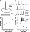
Serotonin (5HT) is a critical modulator of neural circuits that support diverse behaviors and physiological processes, and multiple lines of evidence implicate abnormal serotonergic signaling in psychiatric pathogenesis. The significance of 5HT underscores the importance of elucidating the molecular pathways involved in serotonergic system development, function, and plasticity. However, these mechanisms remain poorly defined, owing largely to the difficulty of accessing 5HT neurons for experimental manipulation. To address this methodological deficiency, we present a transgenic route to selectively alter 5HT neuron gene expression. This approach is based on the ability of a Pet-1 enhancer region to direct reliable 5HT neuron-specific transgene expression in the CNS. Its versatility is illustrated with several transgenic mouse lines, each of which provides a tool for 5HT neuron studies. Two lines allow Cre-mediated recombination at different stages of 5HT neuron development. A third line in which 5HT neurons are marked with yellow fluorescent protein will have numerous applications, including their electrophysiological characterization. To demonstrate this application, we have characterized active and passive membrane properties of midbrain reticular 5HT neurons, which heretofore have not been reported to our knowledge. A fourth line in which Pet-1 loss of function is rescued by expression of a Pet-1 transgene demonstrates biologically relevant levels of transgene expression and offers a route for investigating serotonergic protein structure and function in a behaving animal. These findings establish a straightforward and reliable approach for developing an array of tools for in vivo and in vitro studies of 5HT neurons.
Figures


Similar articles
-
Distinct transcriptomes define rostral and caudal serotonin neurons.J Neurosci. 2010 Jan 13;30(2):670-84. doi: 10.1523/JNEUROSCI.4656-09.2010. J Neurosci. 2010. PMID: 20071532 Free PMC article.
-
A differentially autoregulated Pet-1 enhancer region is a critical target of the transcriptional cascade that governs serotonin neuron development.J Neurosci. 2005 Mar 9;25(10):2628-36. doi: 10.1523/JNEUROSCI.4979-04.2005. J Neurosci. 2005. PMID: 15758173 Free PMC article.
-
Precise pattern of recombination in serotonergic and hypothalamic neurons in a Pdx1-cre transgenic mouse line.J Biomed Sci. 2010 Oct 17;17(1):82. doi: 10.1186/1423-0127-17-82. J Biomed Sci. 2010. PMID: 20950489 Free PMC article.
-
Serotonin neurons and their physiological roles.Arch Histol Cytol. 1989;52 Suppl:113-20. doi: 10.1679/aohc.52.suppl_113. Arch Histol Cytol. 1989. PMID: 2510776 Review.
-
Molecular genetics of the early development of hindbrain serotonergic neurons.Clin Genet. 2005 Dec;68(6):487-94. doi: 10.1111/j.1399-0004.2005.00534.x. Clin Genet. 2005. PMID: 16283875 Review.
Cited by
-
Prospective coding of dorsal raphe reward signals by the orbitofrontal cortex.J Neurosci. 2015 Feb 11;35(6):2717-30. doi: 10.1523/JNEUROSCI.4017-14.2015. J Neurosci. 2015. PMID: 25673861 Free PMC article.
-
An adult-stage transcriptional program for survival of serotonergic connectivity.Cell Rep. 2022 Apr 19;39(3):110711. doi: 10.1016/j.celrep.2022.110711. Cell Rep. 2022. PMID: 35443166 Free PMC article.
-
Postsurgical Latent Pain Sensitization Is Driven by Descending Serotonergic Facilitation and Masked by µ-Opioid Receptor Constitutive Activity in the Rostral Ventromedial Medulla.J Neurosci. 2022 Jul 27;42(30):5870-5881. doi: 10.1523/JNEUROSCI.2038-21.2022. Epub 2022 Jun 14. J Neurosci. 2022. PMID: 35701159 Free PMC article.
-
Adenosine Monophosphate-activated Protein Kinase (AMPK) in serotonin neurons mediates select behaviors during protracted withdrawal from morphine in mice.Behav Brain Res. 2022 Feb 15;419:113688. doi: 10.1016/j.bbr.2021.113688. Epub 2021 Nov 27. Behav Brain Res. 2022. PMID: 34843742 Free PMC article.
-
Adenoviral vectors for highly selective gene expression in central serotonergic neurons reveal quantal characteristics of serotonin release in the rat brain.BMC Biotechnol. 2009 Mar 19;9:23. doi: 10.1186/1472-6750-9-23. BMC Biotechnol. 2009. PMID: 19298646 Free PMC article.
References
-
- Gaspar, P., Cases, O. & Maroteaux, L. (2003) Nat. Rev. Neurosci. 4, 1002-1012. - PubMed
-
- Mason, P. (2001) Annu. Rev. Neurosci. 24, 737-777. - PubMed
-
- Richerson, G. B. (2004) Nat. Rev. Neurosci. 5, 449-461. - PubMed
-
- Sodhi, M. S. & Sanders-Bush, E. (2004) Int. Rev. Neurobiol. 59, 111-174. - PubMed
-
- Barnes, N. M. & Sharp, T. (1999) Neuropharmacology 38, 1083-1152. - PubMed
Publication types
MeSH terms
Substances
Grants and funding
LinkOut - more resources
Full Text Sources
Other Literature Sources
Molecular Biology Databases
Research Materials

