. 2006 Jan;17(1):485-97.
doi: 10.1091/mbc.e05-07-0698. Epub 2005 Oct 26.

The CNA1 histone of the ciliate Tetrahymena thermophila is essential for chromosome segregation in the germline micronucleus

Affiliations

The CNA1 histone of the ciliate Tetrahymena thermophila is essential for chromosome segregation in the germline micronucleus

Marcella D Cervantes et al. Mol Biol Cell. 2006 Jan.

Abstract

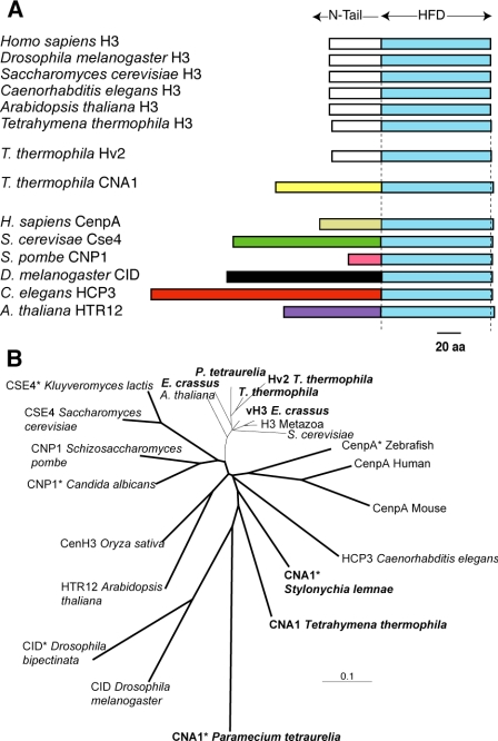
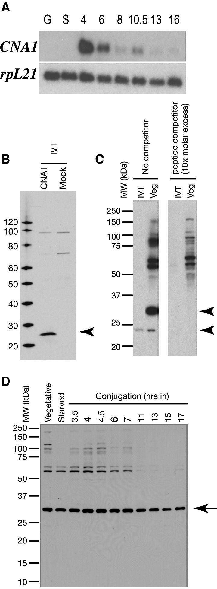
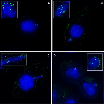
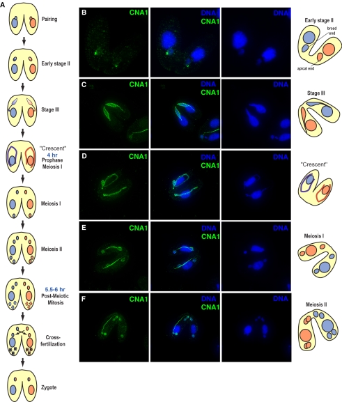
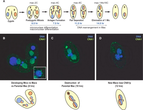
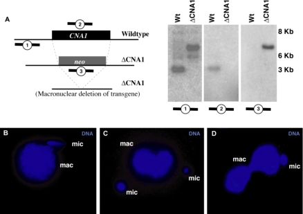
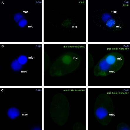
Ciliated protozoans present several features of chromosome segregation that are unique among eukaryotes, including their maintenance of two nuclei: a germline micronucleus, which undergoes conventional mitosis and meiosis, and a somatic macronucleus that divides by an amitotic process. To study ciliate chromosome segregation, we have identified the centromeric histone gene in the Tetrahymena thermophila genome (CNA1). CNA1p specifically localizes to peripheral centromeres in the micronucleus but is absent in the macronucleus during vegetative growth. During meiotic prophase of the micronucleus, when chromosomes are stretched to twice the length of the cell, CNA1p is found localized in punctate spots throughout the length of the chromosomes. As conjugation proceeds, CNA1p appears initially diffuse, but quickly reverts to discrete dots in those nuclei destined to become micronuclei, whereas it remains diffuse and is gradually lost in developing macronuclei. In progeny of germline CNA1 knockouts, we see no defects in macronuclear division or viability of the progeny cells immediately following the knockout. However, within a few divisions, progeny show abnormal mitotic segregation of their micronucleus, with most cells eventually losing their micronucleus entirely. This study reveals a strong dependence of the germline micronucleus on centromeric histones for proper chromosome segregation.


Figures

Figure 1.
Figure 1.
CNA1 is the T. thermophila centromeric histone gene. (A) Schematic comparison of selected canonical histone H3s from eukaryotes showing the conservation of the canonical histones in their histone-fold domains (HFD) and N-terminal tails. In contrast, the centromeric histones do not have a well-conserved N-terminal tail, and this evolves rapidly in both size and sequence. The three histone H3-like genes from T. thermophila are schematized, including the canonical H3, the H3.3 like replacement histone variant hv2 and the centromeric histone CNA1. (B) A neighbor-joining phylogeny of the histone-fold domains of selected canonical and centro-meric histone proteins (bold lines) is shown. Asterisks are used to indicate those proteins that, based on bioinformatic or evolutionary criteria, are thought to be the centromeric histone although this has not been directly tested. This includes putative centromeric histones from other ciliate species, P. tetraurelia and S. lemnae (ciliate histones are shown in bold lettering). In comparison to yeast, vertebrate and plant CenH3s, those from ciliates appear to be rapidly evolving.
Figure 2.
Figure 2.
CNA1 expression. (A) On a Northern blot probed with the CNA1 coding sequence there is no detectable transcript during vegetative growth (G) or starvation (S) but high levels of expression are seen during meiosis (4 h) in conjugation. Expression continues to decrease as conjugation progresses. The rpL21 gene was used as a loading control. (B) Western blot shows that the polyclonal antibody raised to the CNA1 peptide specifically recognizes in vitro-translated (IVT) CNA1p, but no band is detected in the mock expression. (C) The CNA1 antibody recognizes two bands in vegetatively growing T. thermophila cells. The lower band corresponds to the unmodified protein as seen in the in vitro expression and can only be visualized by overexposure, whereas the predominant upper band is ∼5 kDa higher and corresponds to an unknown posttranslationally modified form of CNA1p. Neither band is detected when the antibody staining is competed with a 10-fold molar excess of peptide competitor. (D) CNA1p levels as detected by the Western blot appear stable in vegetative and starved cells, and during conjugation, in stark contrast to the Northern analysis in A.
Figure 3.
Figure 3.
CNA1p localization during mitosis. (A) Immunofluorescence using CNA1p antibody during mitosis shows CNA1p is found only at the centromeres in the micronucleus. During interphase, the centromeres are clustered and peripheral. (B) Centromeres remain peripheral during metaphase when pairs of centromeres align along the middle of the chromatin. (C) The centromeres are on the leading edge of segregating chromosomes in anaphase. (D) By telophase the centromeres are peripheral again. CNA1p staining is shown in green, whereas DNA is stained with DAPI (blue). A weak background staining of basal bodies is seen with the CNA1 antibody, which occasionally outline the cellular boundaries.
Figure 4.
Figure 4.
Early events of conjugation. (A) Conjugation in Tetrahymena is schematized to highlight the major events starting from pairing of cells of different mating types (shown as blue or red) to the formation of the zygote. The diploid micronucleus undergoes meiosis, and one of the four haploid meiotic products is chosen, whereas the other three move to the bottom of the cell and are degraded. The chosen meiotic product divides mitotically and the two cells exchange one haploid nucleus each and fuse to give rise to the zygote (shown as a chimeric red/blue hereafter). (B) During prophase, the micronucleus goes through an egg shape (early stage 2). At this stage, centromeres are clearly visible at the apical end of the nucleus (telomeres have been shown to localize to the basal end at this stage). Micronuclei are then gradually stretched into stage III (C) and eventually the “crescent” shape (D). As the crescent elongates, CNA1p localization can be seen throughout the length of the crescent. (E and F) As meiosis progresses CNA1p remains punctate throughout the micronuclei and also localizes to the meiotic spindle. Distinct centromeres are visible in late anaphase of meiosis I and II. (CNA1p, green; DNA-DAPI, blue). Tubulin staining at these stages of meiosis are presented in Supplementary Information.
Figure 5.
Figure 5.
CNA1 localization during late events in conjugation. (A) Conjugation events leading from zygote formation to progeny cells are schematized. The zygotic nucleus undergoes two rounds of mitosis producing four nuclei (6.5 h), two become micronuclei, whereas two become developing macronuclei (anlagen) and undergo genomic rearrangement and endoduplication. The active parental macronucleus remains in the middle of each cell until the new zygotic macronuclei become active, at ∼7.5 h, and then the parental macronucleus moves to the bottom of the cell and undergoes apoptotic degredation. (B) Eight hours into conjugation CNA1p localization reverts to the peripheral centromeres in the developing micronuclei (inset) but remains diffuse throughout the developing macronuclei. At this time cells remained paired, the parental or old macronucleus has migrated to the bottom of the cell and is beginning apoptotic degradation. (C) By 10 h, CNA1p is concentrated in the old macronucleus that has little DNA remaining. CNA1 localization is decreased in the developing macronuclei, whereas it remains constant at the centromeres in the micronuclei. (D) At the time of genomic rearrangement CNA1p is largely absent from the cell except at the centromeres in the micronuclei, similar to vegetative cells (Figure 3A) (CNA1p, green; DNA-DAPI, blue)
Figure 6.
Figure 6.
Germline knockouts of CNA1 show micronuclear chromosome segregation defects. (A) Southern blots confirming the complete replacement of the CNA1 coding region by the neo cassette in ΔCNA1 cells. Genomic DNA from wild-type and ΔCNA1 cells was digested with XbaI and probed with the upstream genomic region (1), the CNA1 coding sequence (2), or the neo gene (3). The upstream probe detects the wild-type band of 4.1 kb only in wild type. In the ΔCNA1 cells the wild-type band is absent, the most prominent band is the expected 6.2-kb band for the knockout and a small amount of transgene deletion at 5.4 kb was apparent (Yao et al., 2003). The CNA1 gene was absent in the ΔCNA1 cells (middle panel), whereas the neo gene was only detectable in the ΔCNA1 cells (right panel). (B and D) A variety of micronuclear chromosome segregation defects are seen in ΔCNA1 cells, including asymmetric segregation of micronuclear DNA in early anaphase (B) and telophase (C). This asymmetry can lead to cells where the macronuclei divide even though the micronucleus has not (D), whereas typically macronuclear division occurs much later than micronuclear division in wild-type cells.
Figure 7.
Figure 7.
Germline knockouts of CNA1 lead to cells with either large micronuclei or no micronuclei. Micronuclear chromosome segregation defects in ΔCNA1 cells lead to cells that either have abnormally large micronuclei (A and B) or have completely lost their micronucleus (C). Large micronuclei have aberrant CNA1p localization (A). (B) In cells with two large nuclei, large micronuclei were unambiguously identified using antibodies to the micronuclear-specific linker histone δ (Sweet et al., 1996). (C) In cells lacking a cytologically visible micronucleus, no staining with the linker histone δ or CNA1p (unpublished data) was seen. (CNA1p or Linker histone δ, green; DNA-DAPI, blue)

Similar articles

Cited by

References

    1. Ahmad, K., and Henikoff, S. (2002). Histone H3 variants specify modes of chromatin assembly. Proc. Natl. Acad. Sci. USA 99 (Suppl 4), 16477-16484. - PMC - PubMed
    1. Allen, S. L. (2000). Genetic nomenclature rules for Tetrahymena thermophila. Methods Cell Biol. 62, 561-563. - PubMed
    1. Bernhard, D. (1999). Several highly divergent histone H3 genes are present in the hypotrichous ciliate Stylonychia lemnae. FEMS Microbiol. Lett. 175, 45-50. - PubMed
    1. Blomberg, P., Randolph, C., Yao, C. H., and Yao, M. C. (1997). Regulatory sequences for the amplification and replication of the ribosomal DNA minichromosome in Tetrahymena thermophila. Mol. Cell. Biol. 17, 7237-7247. - PMC - PubMed
    1. Blower, M. D., and Karpen, G. H. (2001). The role of Drosophila CID in kinetochore formation, cell-cycle progression and heterochromatin interactions. Nat. Cell Biol. 3, 730-739. - PMC - PubMed

Publication types