. 2005 Dec;11(12):1858-68.
doi: 10.1261/rna.2112705. Epub 2005 Oct 26.

Functional characterization of the SOFA delta ribozyme

Affiliations

Functional characterization of the SOFA delta ribozyme

Lucien Junior Bergeron et al. RNA. 2005 Dec.

Abstract

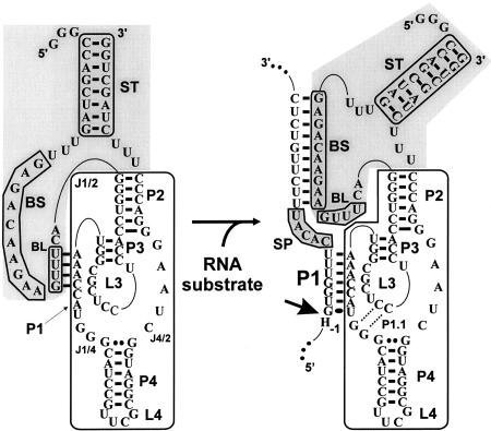
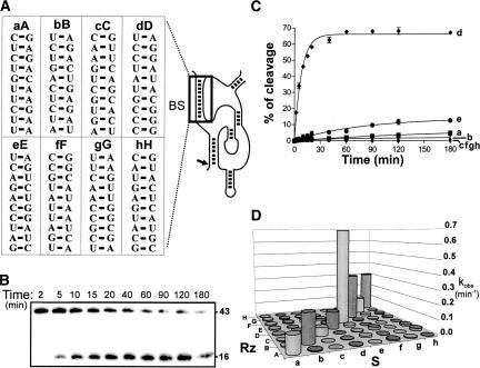
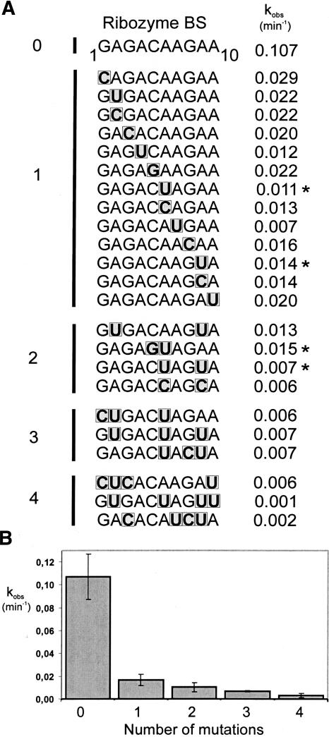
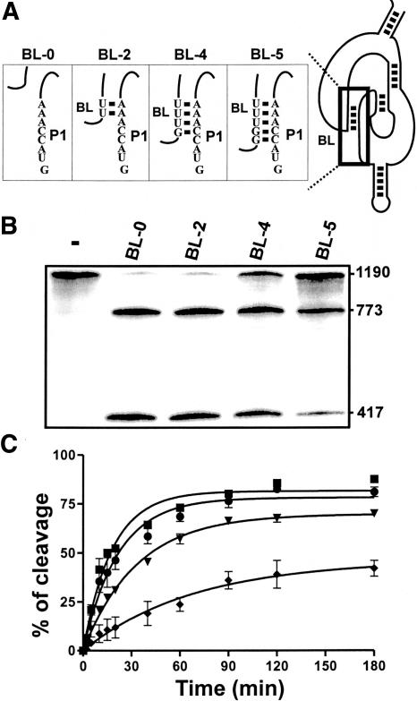
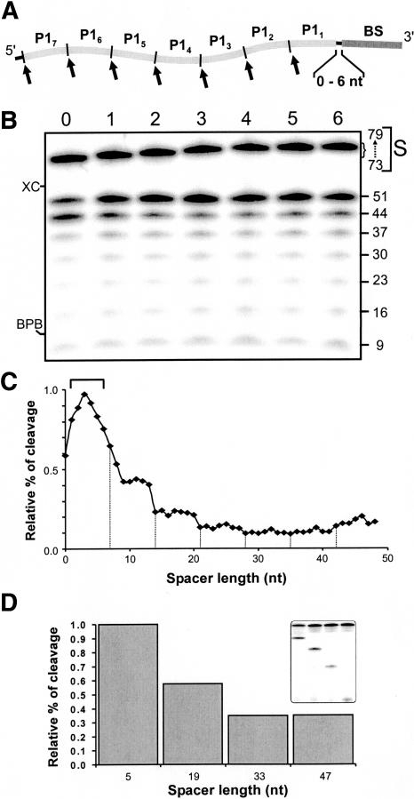
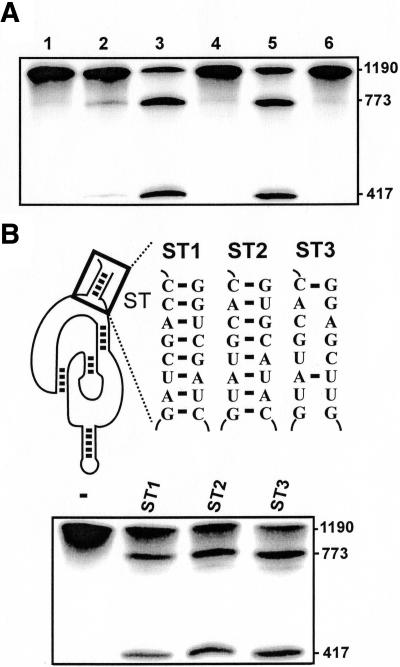
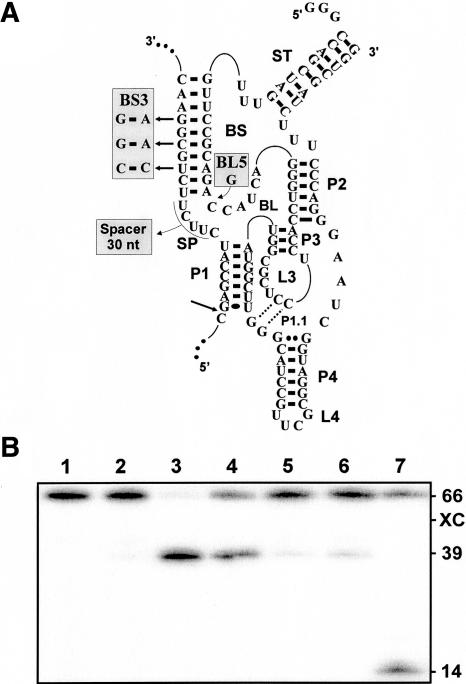
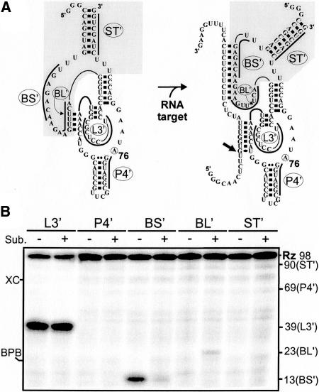
Molecular engineering has led to the development of a novel target-dependent riboswitch that increases deltaribozyme fidelity. This delta ribozyme possesses a specific on/off adapter (SOFA) that switches the cleavage activity from off (a "safety lock") to on solely in the presence of the desired RNA substrate. In this report, we investigate the influence of both the structure and the sequence of each domain of the SOFA module. Analysis of the cleavage activity, using a large collection of substrates and SOFA-ribozyme mutants, together with RNase H probing provided several insights into the nature of the sequence and the optimal design of each domain of the SOFA module. For example, we determined that (1) the optimal size of the blocker sequence, which keeps the ribozyme off in the absence of the substrate, is 4 nucleotides (nt); (2) a single nucleotide difference between the substrate and the biosensor domain, which is responsible for the initial binding of the substrate that subsequently switches the SOFA-ribozyme on, is sufficient to cause non-recognition of the appropriate substrate; (3) the stabilizer, which joins the 5' and 3' ends of the SOFA-ribozyme, plays only a structural role; and (4) the optimal spacer sequence, which serves to separate the binding regions of the biosensor and catalytic domain of the ribozyme on the substrate, is from 1 to 5 nt long. Together, these data should facilitate the design of more efficient SOFA-ribozymes with significant potential for many applications in gene-inactivation systems.


Figures

FIGURE 1.
FIGURE 1.
Schematic representation of both the off and on conformations of SOFA-δRz-303. The gray section indicates the SOFA module; the white section, the original δRz-303. Each investigated segment is boxed in gray: the biosensor sequence (BS), the blocker (BL), the stabilizer (ST), and the spacer (SP). The bold arrow indicates the cleavage site. The H in position − 1 of the substrate represents A, C, or U.
FIGURE 2.
FIGURE 2.
Effect of introducing mutations within the biosensor sequence. (A) Representation of the stems formed between eight pairs of substrate (a–h, left) and ribozyme biosensor (A–H, right) sequences. (B) Autoradiogram of a typical 10% denaturing PAGE gel of a time course experiment performed for the pair Dd. The sizes of the bands are indicated on the right of the gel. (C) Graphical representation of the time course of ribozyme D cleaving each of the substrates (a–h). (D) Histogram of the kobs values for each of the 64 possible pairs.
FIGURE 3.
FIGURE 3.
Fine analysis of the biosensor sequences. (A) Twenty-three biosensor sequence variants were examined for their ability to cleave the short 44-nt HBV-derived substrate. They are clustered on the basis of the number of mutations (zero to four). The mutations are boxed in gray, and the kobs values (in min−1) are indicated on the right. The stars indicate the SOFA-δRz-303 mutants for which the kcat and K M values were determined for the cleavage of a long version of the HBV-derived substrate (1190 nt). (B) Graphical representation of the average values of kobs from at least two independent sets of experiments for each cluster of mutated ribozymes.
FIGURE 4.
FIGURE 4.
Study of the blocker sequence. (A) Representation of the four blocker stems tested. (B) Autoradiogram of a 6% denaturing PAGE of the cleavage assays performed with the SOFA-δRz-303 variants possessing mutated blocker sequences (i.e., BL-X, where X indicates the size of the blocker stem). The reactions were performed under single turnover conditions by using the 1190-HBV substrate. The sizes of the bands are indicated on the right of the gel. The control (−) was performed in the absence of ribozyme. (C) Graphical representation of a kinetic analysis performed for each of the mutants: BL-0, squares; BL-2, circles; BL-4, inverse triangles; and BL-5, diamonds.
FIGURE 5.
FIGURE 5.
Study of the spacer in the substrate. (A) Schematic representation of the design of the substrates used in this experiment. The substrate P1 strand of SOFA-δRz-303 was repeated seven times (P1N, 1–7) within seven substrates possessing spacers of different sizes (0–6 nt). (B) Autoradiogram of a 10% denaturing PAGE of the cleavage assays performed with each of the seven 5′ end labeled substrates. Lanes 0–6 correspond to the different sizes of the spacer sequences (i.e., from 0 to 6 nt). The migrations of the substrates (S) and their sizes, as well as those of the cleavage products, are indicated adjacent to the gel. XC and BPB indicate xylene cyanol and bromophenol blue, respectively. (C) Graphical representation of the relative percentage of cleavage as a function of spacer length. The bracket indicates the optimal length (1–5 nt), and dashed lines separate the observed transitions. (D) Histogram of the relative percentage of cleavage of the substrates possessing spacers of various lengths (5, 19, 33, and 47 nt). The inset shows the autoradiogram of the corresponding 10% denaturing PAGE gel.
FIGURE 6.
FIGURE 6.
Evaluation of the effect of the stabilizer stem on the cleavage activity. (A) Autoradiogram of a 6% denaturing PAGE of cleavage assays of various SOFA-δRz-303 variants. Lane 1 is the incubation of the long HBV-derived substrate (1190 nt) alone, while lane 2 is that in the presence of the original δRz-303. Lanes 3,4 are the cleavage assays performed with SOFA+- and SOFA-δRz-303, including the stabilizer stem, respectively. Lanes 5,6 are the cleavage assays performed with the SOFA+- and SOFA-δRz-303 lacking the stabilizer stem, respectively. The sizes of the bands are indicated adjacent to the gel. (B) On the top are the representations of various stabilizer mutants (SOFA-δRz-303-ST1 to -ST3), while the bottom is the autoradiogram of the denaturing 6% PAGE of the corresponding cleavage assays. The control (−) was performed in the absence of ribozyme.
FIGURE 7.
FIGURE 7.
Evaluation of the predictive value of the determined rules. (A) Schematic representation of SOFA-δRz-HCV. The mutations in both the SOFA-δRz and the substrate are indicated in the shaded boxes. (B) Autoradiogram of a 6% denaturing PAGE gel of the cleavage assays of various SOFA-δRz-HCV variants. Lane 1 is the incubation of the substrate alone (−), while lane 2 is that in the presence of the original δRz-HCV. Lanes 3–5 are the cleavage assays performed with SOFA+-δRz-HCV, SOFA+-δRz-HCV-BL5, and SOFA+-δRz-HCV-BS3, respectively. Lane 6 is the cleavage assay with the SOFA version of δRz-HCV, which possesses an unrelated bio-sensor sequence (i.e., 5′-AUAUAUAUAU-3′). Lane 7 is the cleavage assay performed with the substrate including a 30-nt spacer. The sizes of the bands and the position of the xylen cyanol (XC) are indicated adjacent to the gel.
FIGURE 8.
FIGURE 8.
RNase H probing of SOFA-δRz-303. (A) Schematic representation of the SOFA-ribozyme in both the off and on conformations, respectively. The on conformation is obtained after the addition of the substrate. The bold lines indicate the oligodeoxynucleotides. In these experiments, a C76A (circled residue) mutant that is an inactive version of SOFA-δRz-303 was used. (B) Autoradiogram of an 8% denaturing PAGE of the probing assay. The symbols (−) and (+) indicate the presence or absence, respectively, of the substrate for the probing performed using each oligodeoxynucleotide (L3′, P4′, BS′, BL′, and ST′). The positions of the expected cleavage products, XC and BPB, are indicated adjacent to the gel.
FIGURE 9.
FIGURE 9.
Different conformations for the SOFA module. (A,B) The structures of SOFA+-δRz-Down (SOFA +-δRz-DN, module in the P4 stem) and SOFA+-δRz-Double Binding (SOFA+-δRz-DB, module in both the P2 and P4 stems), respectively. (C) Autoradiogram of a 6% denaturing PAGE gel of cleavage assays performed using these two ribozymes. Lanes 1,2 are the assays performed in the presence of SOFA +-δRz and SOFA-δRz (module in the P2 stem). Lanes 3,4 are the assays performed in the presence of SOFA+-δRz-DN and SOFA-δRz-DN. Lanes 5,6 are the assays in the presence of SOFA+-δRz-DB and SOFA-δRz-DB. All reactions were performed under single turnover conditions. The lengths of the corresponding cleaved bands are indicated beside the gel.

Similar articles

Cited by

References

    1. Al-Anouti, F. and Ananvoranich, S. 2002. Comparative analysis of antisense RNA, double stranded RNA, and delta ribozymemediated gene regulation in toxoplasma gondii. Antisense Nucleic Acid Drug Dev. 12: 275–281. - PubMed
    1. Bagheri, S. and Kashani-Sabet, M. 2004. Ribozymes in the age of molecular therapeutics. Curr. Mol. Med. 4: 489–506. - PubMed
    1. Bergeron, L.J. and Perreault, J.P. 2002. Development and comparison of procedures for the selection of delta ribozyme cleavage sites within the hepatitis B virus. Nucleic Acids Res. 30: 4682–4691. - PMC - PubMed
    1. ———. 2005. Target-dependent on/off switch increases ribozyme fidelity. Nucleic Acids Res. 33: 1240–1248. - PMC - PubMed
    1. Bergeron, L.J., Ouellet, J., and Perreault, J.-P. 2003. Ribozyme-based gene-inactivation systems require a fine comprehension of their substrate specificities: The case of delta ribozyme. Curr. Med. Chem. 10: 2589–2597. - PMC - PubMed

Publication types

MeSH terms