. 2005 Nov;167(5):1207-19.
doi: 10.1016/S0002-9440(10)61209-6.

Conditional ablation of macrophages halts progression of crescentic glomerulonephritis

Affiliations

Conditional ablation of macrophages halts progression of crescentic glomerulonephritis

Jeremy S Duffield et al. Am J Pathol. 2005 Nov.

Abstract

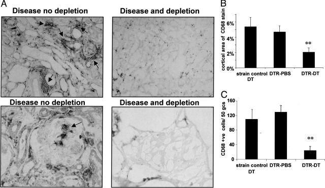
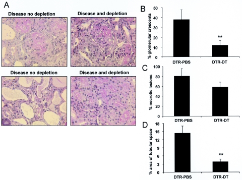
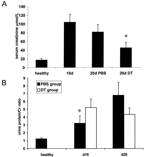
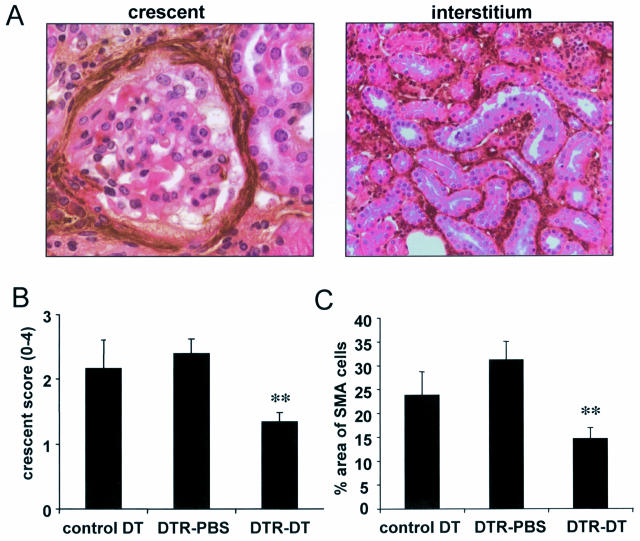
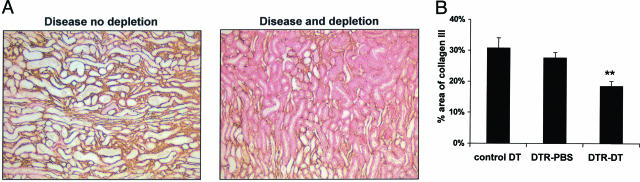
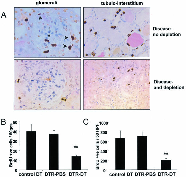
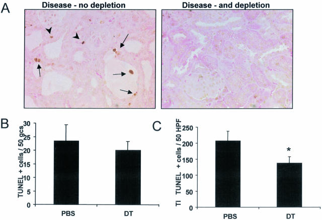
The presence of macrophages in inflamed glomeruli of rat kidney correlates with proliferation and apoptosis of resident glomerular mesangial cells. We assessed the contribution of inflammatory macrophages to progressive renal injury in murine crescentic glomerulonephritis (GN). Using a novel transgenic mouse (CD11b-DTR) in which tissue macrophages can be specifically and selectively ablated by minute injections of diphtheria toxin, we depleted renal inflammatory macrophages through days 15 and 20 of progressive crescentic GN. Macrophage depletion reduced the number of glomerular crescents, improved renal function, and reduced proteinuria. Morphometric analysis of renal tubules and interstitium revealed a marked attenuation of tubular injury that was associated with reduced proliferation and apoptosis of tubular cells. The population of interstitial myofibroblasts decreased after macrophage depletion and interstitial fibrosis also decreased. In the presence of macrophages, interstitial myofibroblasts exhibited increased levels of both proliferation and apoptosis, suggesting that macrophages act to support a population of renal myofibroblasts in a high turnover state and in matrix deposition. Finally, deletion of macrophages reduced CD4 T cells in the diseased kidney. This study demonstrates that macrophages are key effectors of disease progression in crescentic GN, acting to regulate parenchymal cell populations by modulating both cell proliferation and apoptosis.


Figures

Figure 1
Figure 1
Diphtheria toxin successfully depletes glomerular and interstitial macrophages during nephrotoxic nephritis in the CD11b-DTR mouse. A: CD68 immunostaining of tissue sections from mice at 20 days after three doses of diphtheria toxin (DT) commencing at day 15 (right) or control treatment (left). Note areas of intense macrophage infiltration (arrows). Interstitial, crescentic, and intraglomerular macrophages are depleted. B: Morphometric analysis of the whole cortical area indicates that DT significantly reduces the area of tissue occupied by macrophages compared with PBS-treated diseased mice. Note, strain-matched, control diseased mice exhibit an interstitial macrophage infiltrate comparable to PBS-treated CD11b-DTR mice (**P < 0.01). C: The number of intraglomerular macrophages is significantly reduced after DT administration in the CD11b DTR mice compared with PBS-treated diseased mice and strain-matched controls (**P < 0.01).
Figure 2
Figure 2
Histological parameters of renal injury are reduced after conditional macrophage ablation. A: Periodic acid-Schiff-stained sections (×600) from mice at day 20 after three doses of diphtheria toxin (DT) commencing at day 15 (right) or control PBS treatment (left). Glomerular crescents are reduced and tubulointerstitial injury is ameliorated. B: Quantification of the percentage of glomeruli with crescents in DT-treated and PBS-treated diseased CD11b-DTR mice at day 20 (**P < 0.01). C: Quantification of the percentage of glomeruli with necrotic lesions in DT-treated or PBS-treated diseased CD11b-DTR mice. D: Quantitative morphometry of the area of tubular space in the cortex in DT-treated and PBS-treated diseased CD11b-DTR mice (**P < 0.01).
Figure 3
Figure 3
Renal function improves after conditional macrophage ablation. A: Plasma creatinine in healthy CD11b-DTR mice, diseased mice at 15 days, and diseased mice at 20 days after PBS treatment or DT treatment. Note significant reduction in creatinine after macrophage depletion (*P < 0.05). B: Urinary protein to creatinine ratio in healthy CD11b-DTR mice, diseased mice at 15 days, and diseased mice at 20 days after PBS treatment or DT treatment. Note in the PBS-treated group proteinuria increases significantly from day 15 to day 20 (*P < 0.05) whereas in the DT-treated group proteinuria does not increase.
Figure 4
Figure 4
Crescentic and interstitial myofibroblasts are reduced after macrophage ablation. A: Immunolabeling of crescentic and interstitial α-SMA, which is restricted to myofibroblasts. B: Semiquantitative scoring of crescent fibroblasts. Note a significant reduction in DT-treated CD11b-DTR mice but not in strain matched (**P < 0.01). C: Quantitative morphometry of α-SMA-expressing myofibroblasts in the cortical interstitium. Note that myofibroblasts are reduced after DT treatment in the CD11b-DTR mice but not in strain matched control (**P < 0.01).
Figure 5
Figure 5
Interstitial collagen III deposition is reduced after macrophage ablation. A: Immunostaining (×100) for collagen III showing interstitial deposition in diseased mice, which is reduced after macrophage depletion (right). B: Quantitative morphometry of percentage area of cortical collagen III deposition in PBS-treated and DT-treated diseased CD11b-DTR mice and also strain-matched control DT-treated mice (**P < 0.01).
Figure 6
Figure 6
Glomerular and tubulointerstitial proliferation are reduced by macrophage ablation. A: Immunostaining (×600) showing BrdU-positive intraglomerular cells (arrows), periglomerular cells (arrowheads, top left), and (×400) BrdU-positive tubular epithelial and interstitial cells (top right). A reduction in glomerular (bottom left) and tubulointerstitial (bottom right) BrdU-positive cells is seen after macrophage depletion. B: The number of BrdU-positive cells per 50 glomerular cross sections in PBS-treated and DT-treated diseased CD11b-DTR mice and also strain control DT-treated mice (**P < 0.01). C: The number of BrdU-positive cells per 50 HPF of whole kidney in PBS-treated and DT-treated diseased CD11b-DTR mice and also strain control DT-treated mice (**P < 0.01).
Figure 7
Figure 7
Tubulointerstitial apoptosis is reduced by macrophage ablation. A: Immunostaining for apoptotic nuclei using the TUNEL method (×400). Note interstitial apoptosis (arrowhead) and tubular cell apoptosis (arrows) in diseased mice (left) and a reduction in tubulointerstitial apoptosis after macrophage depletion (right). B: The number of intraglomerular apoptotic cells per 50 glomerular cross sections in CD11b-DTR diseased mice treated with PBS or DT. C: The number of tubulointerstitial apoptotic cells per 50 HPF in CD11b-DTR diseased mice treated with PBS or DT (*P < 0.05).

References

    1. Lang R, Lustig M, Francois F, Sellinger M, Plesken H. Apoptosis during macrophage-dependent ocular tissue remodelling. Development. 1994;120:3395–3403. - PubMed
    1. Diez-Roux G, Lang RA. Macrophages induce apoptosis in normal cells in vivo. Development. 1997;124:3633–3638. - PubMed
    1. Diez-Roux G, Argilla M, Makarenkova H, Ko K, Lang RA. Macrophages kill capillary cells in G1 phase of the cell cycle during programmed vascular regression. Development. 1999;126:2141–2147. - PubMed
    1. Duffield JS, Erwig LP, Wei X, Liew FY, Rees AJ, Savill JS. Activated macrophages direct apoptosis and suppress mitosis of mesangial cells. J Immunol. 2000;164:2110–2119. - PubMed
    1. Duffield JS, Ware CF, Ryffel B, Savill J. Suppression by apoptotic cells defines tumor necrosis factor-mediated induction of glomerular mesangial cell apoptosis by activated macrophages. Am J Pathol. 2001;159:1397–1404. - PMC - PubMed

Publication types