. 2005 Nov;167(5):1279-92.
doi: 10.1016/S0002-9440(10)61215-1.

Mouse fetal liver cells in artificial capillary beds in three-dimensional four-compartment bioreactors

Affiliations

Mouse fetal liver cells in artificial capillary beds in three-dimensional four-compartment bioreactors

Satdarshan P S Monga et al. Am J Pathol. 2005 Nov.

Abstract

Bioreactors containing porcine or adult human hepatocytes have been used to sustain acute liver failure patients until liver transplantation. However, prolonged function of adult hepatocytes has not been achieved due to compromised proliferation and viability of adult cells in vitro. We investigated the use of fetal hepatocytes as an alternative cell source in bioreactors. Mouse fetal liver cells from gestational day 17 possessed intermediate differentiation and function based on their molecular profile. When cultured in a three-dimensional four-compartment hollow fiber-based bioreactor for 3 to 5 weeks these cells formed neo-tissues that were characterized comprehensively. Albumin liberation, testosterone metabolism, and P450 induction were demonstrated. Histology showed predominant ribbon-like three-dimensional structures composed of hepatocytes between hollow fibers. High positivity for proliferating cell nuclear antigen and Ki-67 and low positivity for terminal dUTP nick-end labeling indicated robust cell proliferation and survival. Most cells within these ribbon arrangements were albumin-positive. In addition, cells in peripheral zones were simultaneously positive for alpha-fetoprotein, cytokeratin-19, and c-kit, indicating their progenitor phenotype. Mesenchymal components including endothelial, stellate, and smooth muscle cells were also observed. Thus, fetal liver cells can survive, proliferate, differentiate, and function in a three-dimensional perfusion culture system while maintaining a progenitor pool, reflecting an important advance in hepatic tissue engineering.


Figures
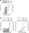
Figure 1
Figure 1
Microarray analysis identifies a multifold increase in gene expression of several genes that reflect synthetic and metabolic function of hepatocytes. A: A graph from a representative gene array analysis demonstrates a notable increase in the expression of many genes such as of albumin, glycerol phosphate dehydrogenase, acetyl coenzyme A dehydrogenase, cytochrome p450 oxidoreductase, apolipoprotein A-1, glycogen storage disease protein 1B, and haptoglobin from E14 to E17 stage (area highlighted), demonstrating acquisition of adult hepatocyte-like synthetic and metabolic functions. B: Left: a similar analysis for several cyp450s (3a13, 3a16, 4a10, 7a1, 2c37, 2d9, 2d10, 2d11) reveals their low expression in E11 and E14 livers with notable up-regulation in E17 liver (highlighted), although these are still low as compared to their respective adult levels. Right: Enlarged highlighted area from the left panel demonstrating a 5- to 100-fold increase in gene expression of these cyp450s in livers from E14 to E17 stage.
Figure 2
Figure 2
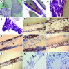
Histology, proliferation, and apoptosis assay of the fetal hepatocytes cultured in bioreactors for 21 days. A: A wet mount of the fixed material under an inverted microscope reveals island of hepatocyte-like cells (arrow) that extend from one hollow fiber (arrowhead) to the other. Inset displays a ribbon-like structure formation. B: H&E staining (low power) shows a few cell-thick ribbon structures (white arrow) that attach to the hollow fibers (arrowhead). Primitive neosinusoidal channels (arrow) can be observed in these structures. C: H&E staining reveals hepatocytes (black arrowhead) within the newly formed tissue. The neosinusoidal spaces (arrow) and duct structure (white arrowhead) can also be observed. D: Another representative H&E staining also reveals ductal structure (white arrowhead) and some flattened cells (arrow) lining the edge of the tissue. E: PCNA immunohistochemistry identifies several proliferating cells (arrow) in ribbon arrangements. Note the negative cells (arrowhead) with flattened morphology at the edges of the tissue. F: High-power view also shows PCNA-positive cells (arrow) in ribbon structures with a few cells in the center and the flattened cell lining staining negative (arrowhead). G: In an adjacent section to E, several Ki-67-positive cells (arrow) are observed in mainly the peripheral area of ribbon-like structures. The cells in the middle part are negative (arrowhead). The flattened cells that line the edge of the tissue are negative (white arrowhead). H: High-power view also emphasizes the differential distribution of Ki-67-positive cells (arrow) toward the periphery of ribbon structures, with predominantly negative (arrowhead) cells toward the center. I: Some areas of ribbon structures show a more uniform PCNA positivity along the entire width with several cells intensely positive for PCNA (arrow). J: An adjacent section of the same area as I also showed Ki-67-positive cells (arrow) occupying the entire width of this part of the ribbon structure. K: Very few apoptotic nuclei (arrow) are observed among the predominantly negative cells (arrowhead) in ribbon-like tissue arrangements by TUNEL staining. L: In the sheet-like arrangement also, <5% of all cells were TUNEL-positive (arrow) with the remaining negatively staining cells (arrowhead). Original magnifications: ×4 (B); ×20 (C–E, G, I–L); ×40 (F, H).
Figure 3
Figure 3
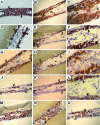
Differentiation of fetal hepatocytes in three-dimensional capillary beds after 21 days in culture. A: Albumin-positive cells (arrowhead) are seen occupying most of the ribbon structures. Negative cells (arrow) are flattened cells that line the edge of the tissue. B: Another similar ribbon-like structure showing most cells to be albumin-positive. C: High power of B reveals albumin-positive hepatocytes (black arrowhead) several of them arranged being traversed by neosinusoidal structures (white arrowhead) and lined at either edge by flattened albumin-negative cells (white arrow). D: α-FP-positive cells (black arrowhead) are observed toward either peripheral zone in the ribbon structures. The cells in the middle zone (black arrow) and the flattened cell lining at the edge (white arrow) stains negative for α-FP. E: An adjacent section to B displays a subset of albumin-positive cells, mostly toward either periphery (arrowhead) to be α-FP-positive. Again the flattened cells toward the edges are negative. F: High power of E clearly shows α-FP-positive cells toward the periphery (arrowhead) and the negative cells toward the middle of the ribbon-like structures (black arrow). The flattened endothelial cells are α-FP-negative (white arrow). G: C-Kit-positive cells (arrowhead) are observed toward the periphery with negatively staining hepatocytes (black arrow) in the central region and flattened cells at the edges (white arrow). H: An adjacent section to B and E also demonstrates c-kit-positive cells toward the periphery (arrowhead) and negative cells in the middle zone (black arrow) and at the edges (white arrow). I: High power shows intensely staining c-kit-positive cells toward the side of ribbons (arrowhead) surrounding the negative hepatocytes in the middle (black arrow). Flattened cells at edges were c-kit-negative (white arrow). J: CK-19-positive cells (arrowhead) are observed at the peripheral zones in ribbon structures whereas the negative cells (black arrow) occupy the middle zone. Again the flattened cells at the edge were negative (arrowhead). K: An adjacent section to B, E, or H, also shows predominant CK-19-positive cells toward the side (arrowhead) whereas cells in the middle (black arrow) or edges (white arrow) are negative. L: A high power of this area reveals hepatocyte-like cells at the periphery to be CK-19-positive (arrowhead) and in the central region to be negative (arrow). M: Minority of ribbon-like structures displayed most cells (almost entire thickness) to be α-FP-positive (arrowhead) with a few negatively staining cells (black arrow). Flattened cells at edges were always negative (white arrow). N: An adjacent section to M shows the same cells to be also positive for c-kit (arrowhead) throughout the thickness of such ribbon structures. Again a few cells were negative (black arrow) and the cells at edges were negative as well (white arrow). O: Another adjacent section to M and N displaying similar distribution of CK-19-positive cells (arrowhead) with a few negatively staining cells interspersed in between (black arrow) or observed at edges (white arrow). Original magnifications: ×20 (A, B, D, E, G, J, K, M–O); ×40 (C, F, I); ×60 (L).
Figure 4
Figure 4
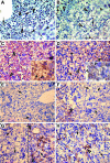
Bioreactor cultures also display mesenchymal differentiation. A: Isolectin-staining positive cells, with flattened morphology (arrowhead), are observed at the edges of this sheet-like arrangement of cells that is composed of many negatively staining epithelial cells (arrow). B: Desmin staining reveals an area predominantly composed of nonepithelial cells to have several desmin-positive stellate cells (arrowhead). Most other cells were negative (arrow). C: A high-power view of D reveals a large stellate cell with punctate desmin staining (arrowhead). D: Several other areas in ribbon formations were devoid of desmin-positive cells as seen in low magnification in left panel and high power in right panel that display mainly epithelial cells (arrow). E: α-SMA staining shows certain areas of the culture to contain myofibroblasts (arrowhead) interspersed between negatively staining epithelial cells (arrow). F: Most areas in ribbons had only minimal α-SMA positivity (inset and arrowhead) whereas most other epithelial cells were clearly negative (arrow). G: Vimentin staining was negative in most of the ribbon arrangements (arrow). H: High-power view also shows predominantly negatively staining cells for vimentin (arrow) with very few faintly positive cells that have hepatocyte morphology (arrowhead) and might represent EMT. I: Only a few areas showed more numbers of vimentin-positive cells (arrowhead and inset at left). Other areas showed larger stellate cells to be positive for vimentin as well (right). Original magnifications: × 20 (A, B, F, G); × 40 (C, E, inset in F, H).
Figure 5
Figure 5
Proliferation and differentiation in E17 mice livers. A: PCNA-positive cells (arrow) constitute ∼30% of E17 liver as seen in this representative section. Most other cells are PCNA-negative (arrowhead). A higher magnification inset shows positive (arrow) and negative cells (arrowhead) with greater precision. B: A few (<15%) cells in E17 liver were Ki-67-positive (arrow). The predominant cell population was Ki-67-negative (arrowhead). Inset shows positively (arrow) and negatively staining cells (arrowhead) more clearly. C: A representative section shows almost all cells to be albumin-positive (arrowhead) in an E17 liver. A higher magnification inset shows a hematopoietic cell staining negative (arrow) as compared to other cells that are albumin-positive (arrowheads). D: Approximately half of the cells in E17 livers were α-FP-positive (arrowhead) whereas the remaining cells were negative (arrow). A higher magnification shows α-FP-positive (arrowheads) and -negative (arrows) cells. E: A representative section from an E17 liver shows all cells negative for c-kit (arrow). F: Another representative section shows a small group of c-kit-positive cells (arrowhead) within predominantly negative cells (arrow). G: Left panel represents all cells negative for CK-19 (arrow) in a representative E17 liver section. The right panel represents isolated CK-19 cells (black arrowhead) representing progenitor cell fraction. Although most cells are CK-19-negative (arrow), primitive portal plates (white arrowhead) are CK-19-positive. H: Another representative section from E17 liver demonstrates isolated CK-19-positive cells (black arrowhead), portal plates (white arrowhead) among mostly CK-19-negative cells (arrow). Original magnifications: ×40.
Figure 6
Figure 6
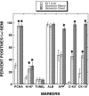
Comparison of cell populations after culture of E17 liver cells in bioreactors for 21 days. A significant increase in the number of PCNA- and Ki-67-positive cells after the culture of E17 cells indicates robust cell proliferation (P < 0.01). No adverse increases in TUNEL-positive cell numbers were observed after the culture of E17 liver cells indicating apoptosis within physiological limits and good cell viability of fetal hepatocytes in the bioreactors. Although the numbers of albumin-positive cells remained unaltered, a significant increase was observed in the numbers of α-FP-, c-kit-, and CK-19-positive cells (P < 0.05). Although α-FP positivity was comparable to the E17 livers in the ribbon-like structures, tissue islands were composed of either all positive or all negative cells (*P < 0.05; +cells are counted in tissue islands that were composed of all positive cells for α-FP, c-kit, and CK-19).
Figure 7
Figure 7
Long-term experiment to measure albumin liberation (triangles, left axis) and P450 activity (CYP3A) (bars, right axis). Albumin liberation increased for the first week, then maintained a relatively stable rate for the remaining 4 weeks. Testosterone metabolism to 6β-hydroxytestosterone doubled after pregnenolone carbonitrile induction.

References

    1. Patzer IJ, Lopez RC, Zhu Y, Wang ZF, Mazariegos GV, Fung JJ. Bioartificial liver assist devices in support of patients with liver failure. Hepatobiliary Pancreat Dis Int. 2002;1:18–25. - PubMed
    1. Gerlach JC. Development of a hybrid liver support system: a review. Int J Artif Organs. 1996;19:645–654. - PubMed
    1. Rudow DL, Russo MW, Hafliger S, Emond JC, Brown RS., Jr Clinical and ethnic differences in candidates listed for liver transplantation with and without potential living donors. Liver Transpl. 2003;9:254–259. - PubMed
    1. Dowling DJ, Mutimer DJ. Artificial liver support in acute liver failure. Eur J Gastroenterol Hepatol. 1999;11:991–996. - PubMed
    1. Monga A, Jain SK. Artificial liver support systems. J Assoc Physicians India. 1999;47:1092–1099. - PubMed

Publication types

MeSH terms

LinkOut - more resources