

Matched skin and sentinel lymph node samples of melanoma patients reveal exclusive migration of mature dendritic cells
- PMID: 16251414
- PMCID: PMC1603792
- DOI: 10.1016/S0002-9440(10)61217-5
Matched skin and sentinel lymph node samples of melanoma patients reveal exclusive migration of mature dendritic cells
Abstract
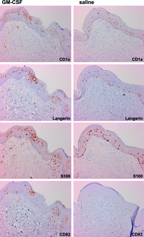
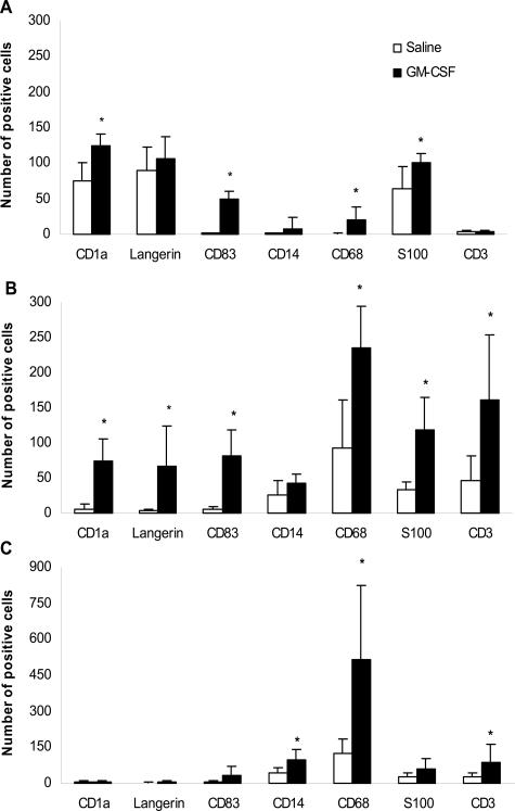
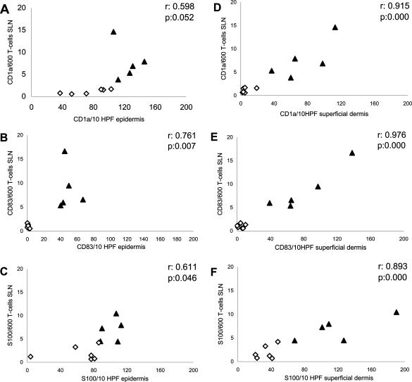
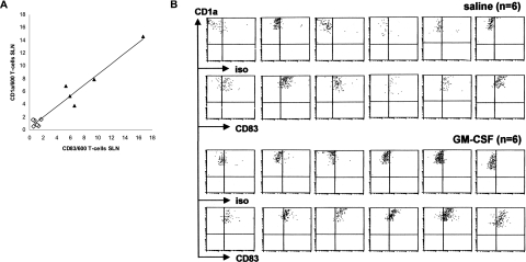
Mature and immature myeloid dendritic cells (DCs) are thought to differentially modulate T-cell responses in secondary lymphoid tissues. Although mature DCs are believed to induce T-cell activation under proinflammatory conditions, immature DCs are believed to maintain a state of T-cell tolerance under steady state conditions. However, little is known about the actual activation state of human DCs under these different conditions. Here, we compare the frequency and activation state of human DCs between matched skin and sentinel lymph node (SLN) samples, after intradermal administration of either granulocyte/macrophage colony-stimulating factor (GM-CSF) or saline, at the excision site of stage I primary melanoma. Although DCs remained immature (CD1a+CD83-) and mostly situated in the epidermis of the saline-injected skin (fully consistent with a quiescent steady state), mature (CD1a+CD83+) DC frequencies significantly increased in the GM-CSF-injected skin and correlated with the number of mature DCs in the SLN, indicative of increased DC migration. Interestingly, irrespective of GM-CSF or saline administration, all CD1a+ myeloid DCs in the SLN were phenotypically mature (ie, CD83+). These data are indicative of migration of small numbers of phenotypically mature DCs to lymph nodes under steady state conditions.
Figures
References
-
- Smith CH, Allen MH, Groves RW, Barker JN. Effect of granulocyte macrophage-colony stimulating factor on Langerhans cells in normal and healthy atopic subjects. Br J Dermatol. 1998;139:239–246. - PubMed
-
- Caux C, Dezutter-Dambuyant C, Schmitt D, Banchereau J. GM-CSF and TNF-alpha cooperate in the generation of dendritic Langerhans cells. Nature. 1992;360:258–261. - PubMed
-
- Steinman RM, Hawiger D, Nussenzweig MC. Tolerogenic dendritic cells. Annu Rev Immunol. 2003;21:685–711. - PubMed
-
- Henri S, Vremec D, Kamath A, Waithman J, Williams S, Benoist C, Burnham K, Saeland S, Handman E, Shortman K. The dendritic cell populations of mouse lymph nodes. J Immunol. 2001;167:741–748. - PubMed
Publication types
MeSH terms
Substances
LinkOut - more resources
Full Text Sources
Other Literature Sources
Medical
