. 2005 Nov;167(5):1309-20.
doi: 10.1016/S0002-9440(10)61218-7.

Bacillus anthracis edema toxin causes extensive tissue lesions and rapid lethality in mice

Affiliations

Bacillus anthracis edema toxin causes extensive tissue lesions and rapid lethality in mice

Aaron M Firoved et al. Am J Pathol. 2005 Nov.

Abstract

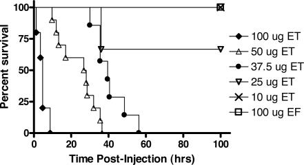
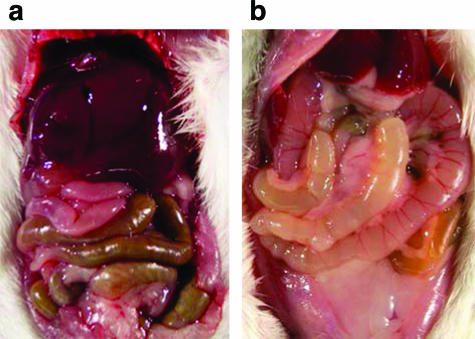
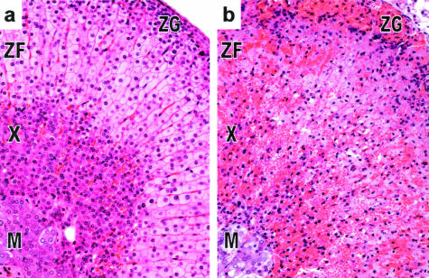
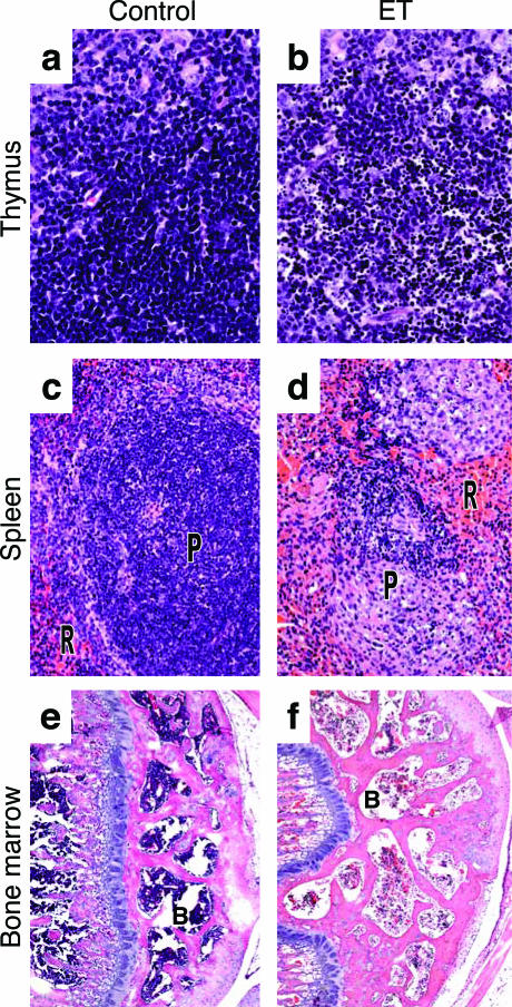
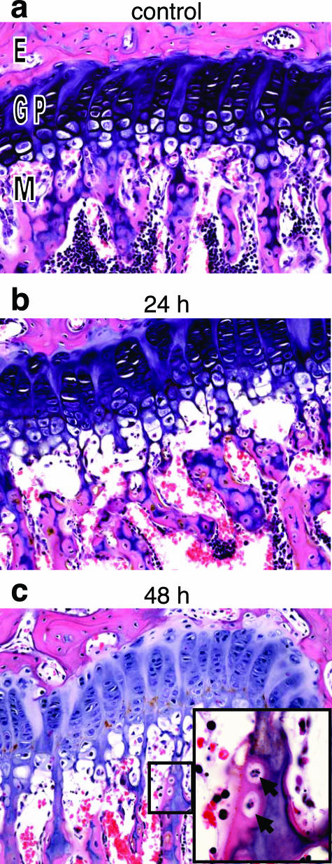
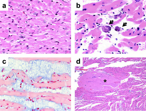
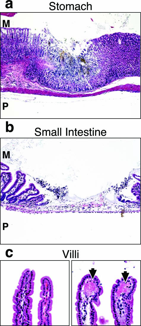
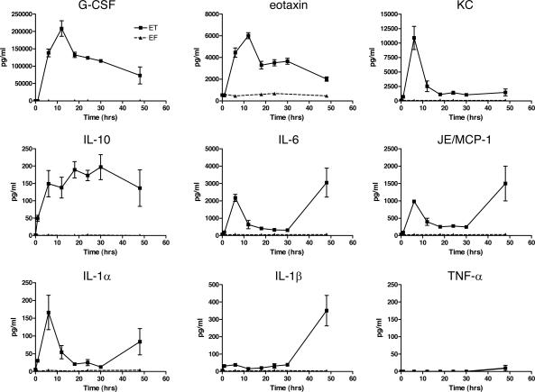
Bacillus anthracis edema toxin (ET), an adenylyl cyclase, is an important virulence factor that contributes to anthrax disease. The role of ET in anthrax pathogenesis is, however, poorly understood. Previous studies using crude toxin preparations associated ET with subcutaneous edema, and ET-deficient strains of B. anthracis showed a reduction in virulence. We report the first comprehensive study of ET-induced pathology in an animal model. Highly purified ET caused death in BALB/cJ mice at lower doses and more rapidly than previously seen with the other major B. anthracis virulence factor, lethal toxin. Observations of gross pathology showed intestinal intralumenal fluid accumulation followed by focal hemorrhaging of the ileum and adrenal glands. Histopathological analyses of timed tissue harvests revealed lesions in several tissues including adrenal glands, lymphoid organs, bone, bone marrow, gastrointestinal mucosa, heart, and kidneys. Concomitant blood chemistry analyses supported the induction of tissue damage. Several cytokines increased after ET administration, including granulocyte colony-stimulating factor, eotaxin, keratinocyte-derived cytokine, MCP-1/JE, interleukin-6, interleukin-10, and interleukin-1beta. Physiological measurements also revealed a concurrent hypotension and bradycardia. These studies detail the extensive pathological lesions caused by ET and suggest that it causes death due to multiorgan failure.


Figures

Figure 1
Figure 1
ET lethality in BALB/cJ mice. ET-treated female BALB/cJ mice at doses of 100 μg (n = 5), 50 μg (n = 10), 37.5 μg (n = 7), 25 μg (n = 6), and 10 μg (n = 6), were compared to animals treated with 100 μg of EF alone (control). All injections were intravenous in 100-μl volume. All differences in survival curves when compared against each other were statistically significant (P < 0.05) by the log-rank test except for the comparison between the 25 μg of ET dose and 100 μg of EF dose (P = 0.1573).
Figure 2
Figure 2
ET induces marked intestinal dilatation. Mice were injected intravenously with 37.5 μg of EF alone (control) (a) or ET (b) and euthanized 12 hours later by CO2 inhalation and immediately necropsied.
Figure 3
Figure 3
ET causes adrenal gland lesions. Histopathological evaluation of adrenal glands at 12 hours in EF-treated (control) (a) and ET-treated (b) animals revealed necrosis and hemorrhage of the zona fasciculata (ZF) of the adrenal cortex induced by ET. The X-Zone (X) and zona glomerosa (ZG) were moderately necrotic whereas the medulla (M) was minimally affected by ET. Original magnifications, ×20.
Figure 4
Figure 4
Histopathological analysis of lymphoid tissues. Mice were injected intravenously with 37.5 μg of EF alone (control), PBS, or 37.5 μg of ET before histopathological analysis of lymphoid tissues. a: Thymus from EF-treated (36 hours) control. b: Nuclear fragmentation of lymphocytes in the thymus of ET-treated animals (36 hours). c: Red pulp (R) and periarteriolar lymphoid sheath (P) of spleen of PBS-treated (48 hours) control. d: Necrotic splenic periarterial lymphoid sheath in ET-treated (48 hours) animals. e: Bone marrow (B) in PBS-treated (48 hours) control. f: Depletion of hematopoietic cells of the bone marrow in ET-treated mice (48 hours). Original magnifications: ×40 (a, b); ×20 (c, d); ×5 (e, f).
Figure 5
Figure 5
Bone degeneration after ET administration. Histopathological analysis of femur after intravenous injection with 37.5 μg of EF (control), PBS, or 37.5 μg of ET. a: Growth plate from femur of PBS control (24 hours). b and c: Loss of osteoblasts and osteocytes of the metaphyseal bone marrow at 24 (b) and 48 (c) hours after ET treatment. Inset: Lacunae containing necrotic osteocytes (solid arrows). E, epiphysis; GP, growth plate; M, metaphysis. Original magnifications: ×20; ×100 (inset).
Figure 6
Figure 6
ET-induced cardiac damage. Histopathological analysis of cardiac tissues after intravenous injection with 37.5 μg of EF, PBS, or 37.5 μg of ET. a: Cardiomyocytes from PBS-treated (48 hours) control. b: Cardiomyocyte degeneration in ET-treated mice (arrows, 48 hours). c: Interstitial myxoid deposits in heart tissue stained with Alcian blue (48 hours). d: Thrombus formation at 12 hours after ET-treatment (asterisk). Original magnifications: ×40 (a–c); ×5 (d).
Figure 7
Figure 7
Fibrin deposition and mucosal degeneration of GI tract caused by ET. Histopathology of GI tract mucosa after intravenous injection with 37.5 μg of EF, PBS, or 37.5 μg of ET. a: Areas of focal necrosis surrounded by normal tissue in the stomach mucosa of ET-treated mice (36 hours). b: Focal necrosis of the intestine 24 hours after ET treatment. c: Fibrin deposition in the villus tips in ET-treated animals (24 hours; right, arrows) as compared to normal villi from control animals (24 hours, left). M, mucosal surface; P, peritoneal cavity. Original magnifications: ×5 (a); ×10 (b); ×20 (c).
Figure 8
Figure 8
ET-induced cytokine response. Measurements of serum cytokine levels after intravenous injection of 37.5 μg of ET or EF (control). Results represent averages from duplicate measurements done on three pools of sera, each deriving from two mice except for the 48-hour ET-treated animals in which duplicate measurements were done on sera drawn from three individual animals.
Figure 9
Figure 9
Cardiovascular response to ET exposure. A: Mean heart rate (bpm, open boxes) and arterial blood pressure response (mmHg, filled boxes) after intraperitoneal ET (100 μg) injection (n = 4). The dashed line represents the time of injection. Both heart and blood pressure declined simultaneously. B: Representative electrocardiogram tracings at baseline and timed intervals after exposure to ET. QRS widening and heart block can be seen at 4 hours after ET exposure. C: Constant mean arterial blood pressure after intraperitoneal injection of 100 μg of mutant E346R ET (n = 2, open boxes) as compared to native ET (n = 4, filled boxes) plotted in A.

References

    1. Turnbull PC. Introduction: anthrax history, disease and ecology. Curr Top Microbiol Immunol. 2002;271:1–19. - PubMed
    1. Jernigan JA, Stephens DS, Ashford DA, Omenaca C, Topiel MS, Galbraith M, Tapper M, Fisk TL, Zaki S, Popovic T, Meyer RF, Quinn CP, Harper SA, Fridkin SK, Sejvar JJ, Shepard CW, McConnell M, Guarner J, Shieh WJ, Malecki JM, Gerberding JL, Hughes JM, Perkins BA. Bioterrorism-related inhalational anthrax: the first 10 cases reported in the United States. Emerg Infect Dis. 2001;7:933–944. - PMC - PubMed
    1. Kuehnert MJ, Doyle TJ, Hill HA, Bridges CB, Jernigan JA, Dull PM, Reissman DB, Ashford DA, Jernigan DB. Clinical features that discriminate inhalational anthrax from other acute respiratory illnesses. Clin Infect Dis. 2003;36:328–336. - PubMed
    1. Guarner J, Jernigan JA, Shieh WJ, Tatti K, Flannagan LM, Stephens DS, Popovic T, Ashford DA, Perkins BA, Zaki SR. Pathology and pathogenesis of bioterrorism-related inhalational anthrax. Am J Pathol. 2003;163:701–709. - PMC - PubMed
    1. Barakat LA, Quentzel HL, Jernigan JA, Kirschke DL, Griffith K, Spear SM, Kelley K, Barden D, Mayo D, Stephens DS, Popovic T, Marston C, Zaki SR, Guarner J, Shieh WJ, Carver HW, Meyer RF, Swerdlow DL, Mast EE, Hadler JL. Fatal inhalational anthrax in a 94-year-old Connecticut woman. JAMA. 2002;287:863–868. - PubMed

Publication types

MeSH terms