Comparative Study
. 2005 Oct 26;25(43):9883-92.
doi: 10.1523/JNEUROSCI.1531-05.2005.

Role of hippocampal Cav1.2 Ca2+ channels in NMDA receptor-independent synaptic plasticity and spatial memory

Affiliations
Comparative Study

Role of hippocampal Cav1.2 Ca2+ channels in NMDA receptor-independent synaptic plasticity and spatial memory

Sven Moosmang et al. J Neurosci. .

Abstract

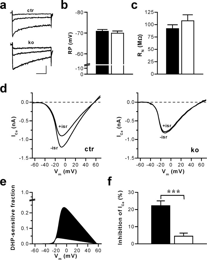
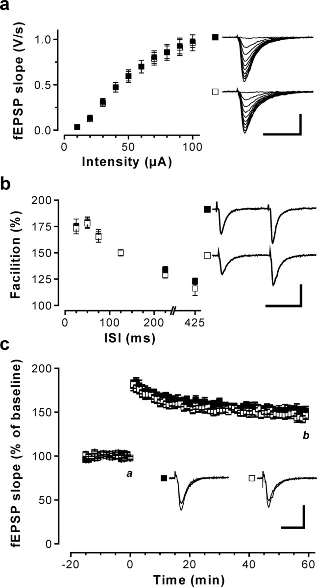
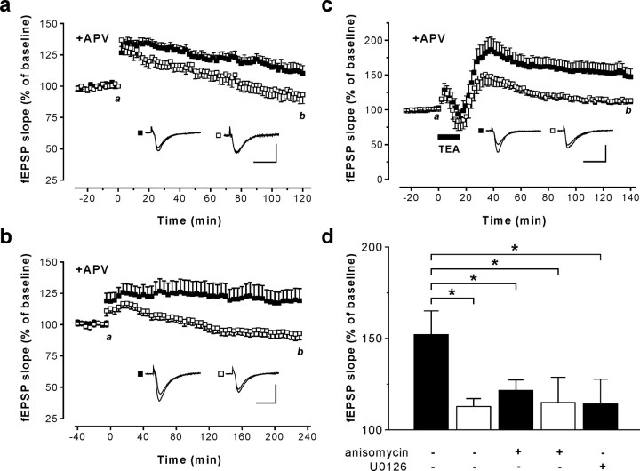
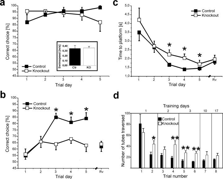
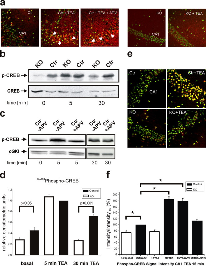
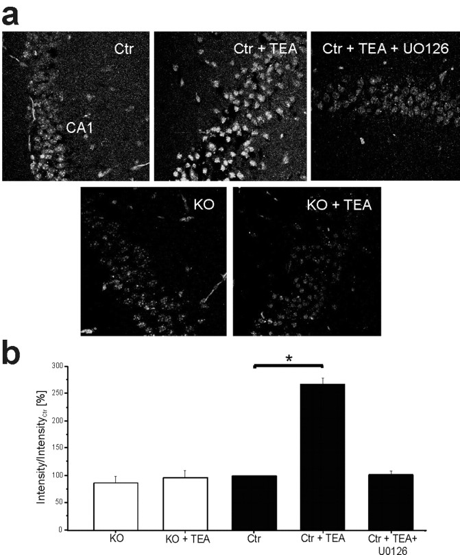
Current knowledge about the molecular mechanisms of NMDA receptor (NMDAR)-independent long-term potentiation (LTP) in the hippocampus and its function for memory formation in the behaving animal is limited. NMDAR-independent LTP in the CA1 region is thought to require activity of postsynaptic L-type voltage-dependent Ca2+ channels (Cav1.x), but the underlying channel isoform remains unknown. We evaluated the function of the Cav1.2 L-type Ca2+ channel for spatial learning, synaptic plasticity, and triggering of learning-associated biochemical processes using a mouse line with an inactivation of the CACNA1C (Cav1.2) gene in the hippocampus and neocortex (Cav1.2(HCKO)). This model shows (1) a selective loss of protein synthesis-dependent NMDAR-independent Schaffer collateral/CA1 late-phase LTP (L-LTP), (2) a severe impairment of hippocampus-dependent spatial memory, and (3) decreased activation of the mitogen-activated protein kinase (MAPK) pathway and reduced cAMP response element (CRE)-dependent transcription in CA1 pyramidal neurons. Our results provide strong evidence for a role of L-type Ca2+ channel-dependent, NMDAR-independent hippocampal L-LTP in the formation of spatial memory in the behaving animal and for a function of the MAPK/CREB (CRE-binding protein) signaling cascade in linking Cav1.2 channel-mediated Ca2+ influx to either process.


Figures

Figure 1.
Figure 1.
CA1 pyramidal cells of mice with an inactivation of the CACNA1C gene (in Cav1.2HCKO) in the hippocampus display a nearly complete loss of L-type Ca2+ currents without changes in RP and RN. a, Ca2+ channel currents evoked by voltage-clamp steps in pyramidal neurons of hippocampal slices from control (ctr) and Cav1.2HCKO (ko) mice. The traces illustrated correspond to test potentials of -30, -20, and -10 mV. The holding potential was -65 mV, and 2 mm Ca2+ was used as a charge carrier. Calibration: 50 ms, 100 pA. b, Average RP measured in CA1 neurons of hippocampal slices from control (▪) (n = 13 cells from 6 mice) and Cav1.2HCKO (□) (n = 10 cells from 6 mice) mice in the current-clamp mode. Means + SEM are shown. c, Average RN of hippocampal CA1 neurons from control (▪) (n = 8 cells from 5 mice) and Cav1.2HCKO (□) (n = 6 cells, 5 mice) mice determined by injecting hyperpolarizing currents from a membrane potential of -70 mV. Means + SEM are shown. d, Representative examples of whole-cell Ca2+ currents (ICa) evoked by voltage-clamp ramps from -80 to +80 mV (0.5 ms/mV) in hippocampal CA1 neurons from ctr and ko mice before (-isr) and after (+isr) application of the L-type channel blocker isradipine (10 μm). The holding potential was -65 mV, and 2 mm Ca2+ was used as a charge carrier. e, Fraction of the total voltage-dependent Ca2+ inward current blocked by isradipine for the representative examples shown in d. Fitted I-V curves of the DHP-sensitive current component in control (filled in black) and Cav1.2HCKO (filled in white) mice, normalized to the corresponding peak inward current in the absence of isradipine (for details on fit function, see Materials and Methods), are represented. f, Average component of the total voltage-dependent Ca2+ current block by isradipine. The reduction of the peak inward current in hippocampal CA1 neurons from control (▪) (n = 14 cells from 6 mice) and Cav1.2HCKO (□) (n = 9 from 5 mice) mice, shown in percentage, is represented. Data are means ± SEM (***p < 0.001).
Figure 2.
Figure 2.
Basal synaptic transmission and NMDAR-dependent synaptic plasticity in the hippocampal CA1 region of Cav1.2HCKO mice. fEPSPs in response to stimulation of the Schaffer collaterals were recorded in slices from control (▪) and Cav1.2HCKO (□) mice. a, Input-output relationship curves obtained from control (37 slices from 16 mice) and Cav1.2HCKO mice (41 slices from 21 mice) were not different. Representative recordings are shown on the right (calibration: 20 ms, 1 mV). b, Short-term plasticity was assessed by measuring PPF at interstimulus intervals (ISIs) of 25, 50, 75, 125, 225, and 425 ms. PPF was not altered in control (n = 27 slices from 12 mice) compared with Cav1.2HCKO mice (n = 25 slices from 13 mice). Representative PPF recordings for the 75 ms interval are shown on the right (calibration: 50 ms, 1 mV). c, NMDAR-dependent Schaffer collateral/CA1 LTP was induced by theta-burst stimulation (12 times 4 pulses at 100 Hz; 200 ms pause) applied at time 0. The time course of the fEPSP slope (mean + SEM) was obtained from control (8 slices from 3 mice) and Cav1.2HCKO (9 slices from 4 mice) slices. Representative fEPSPs recorded at the times indicated (a, b) are shown in the corresponding inset. Calibration: 20 ms, 1 mV. Error bars indicate SE.
Figure 3.
Figure 3.
NMDAR-independent LTP in the hippocampal CA1 region of Cav1.2HCKO mice. fEPSPs in response to stimulation of the Schaffer collaterals were recorded in slices from control (▪) and Cav1.2HCKO (□) mice. a, LTP was induced at time 0 by a 200 Hz tetanic stimulation with the NMDAR antagonist APV (50 μm) present throughout the experiment. The time course of the fEPSP slope represents mean + SEM of experiments in eight slices from five mice for each genotype. Representative fEPSPs recorded at the times indicated (a, b) are shown in the corresponding inset. b, LTP was induced at time 0 by multiple strong 100 Hz tetani with the NMDAR antagonist APV (50 μm) present throughout the experiment. The time course of the fEPSP slope represents the mean + SEM in control (10 slices from 6 mice) and Cav1.2HCKO (14 slices from 6 mice) slices. Representative fEPSPs recorded at the times indicated (a, b) are shown in the corresponding inset. c, LTPK was induced by superfusing hippocampal slices with aCSF containing 25 mm potassium channel blocker TEA for 15 min (indicated by the bar). The NMDAR antagonist, APV (50 μm), was present throughout the experiment. The time course of the fEPSP slope represents the mean ± SEM in control (15 slices from 12 mice) and Cav1.2HCKO mice (11 slices from 7 mice). Representative fEPSPs recorded at the times indicated (a, b) are shown in the corresponding inset. d, Effect of the protein synthesis inhibitor anisomycin (20 μm) and the MEK1/2 inhibitor U0126 (40 μm) on L-LTPK in control and Cav1.2HCKO mice. ▪, The magnitude of LTPK 2 h after treatment with TEA in control mice in normal aCSF, in the presence of anisomycin (n = 8 slices from 5 mice) and U0126 (n = 7 slices from 5 mice) is illustrated. □, The magnitude of LTPK 2 h after treatment with TEA in control mice in normal aCSF and in the presence of anisomycin (n = 11 slices from 7 mice), respectively, is illustrated. *Significant difference (p < 0.05). Calibrations: 20 ms, 1 mV. Error bars indicate SE.
Figure 4.
Figure 4.
Spatial memory defects in Cav1.2HCKO mice. a, Correct-choice rate of mice trained to find the correct platform in a visual acuity task by using the only a visible landmark in a dimly lit room. There were no differences between the Cav1.2HCKO (□) animals (n = 7) and the control (▪) mice (n = 7) in the percentage of correct choices during the trial days (10 trials per day). Inset, Swim speed. Cav1.2HCKO (□) animals (n = 10) present the same swim-speed rate (meters/second) compared with control (▪) mice (n = 10). Data are expressed as mean + SEM. b, c, The performance of Cav1.2HCKO mice in the discriminatory water-maze task. b, The graph represents the correct-choice rate of mice trained to find the correct platform in a discriminatory water maze by using the distal cues surrounding it. The Cav1.2HCKO (□) animals (n = 10) display a significantly (p < 0.05) lower correct-choice rate from day 3 (10 trials per day) than the control (▪) mice (n = 10). During a control reversal trial (Rv), the percentage of correct choices made by control mice dropped by a similar extent to values near chance level, indicating that spatial searching strategies wereused. c, The graph represents the escape latencies of mice that found the correct platform. The Cav1.2HCKO (□) animals (n = 10) displayed a longer latency, starting from day 3 (10 trials per day) than the control (▪) mice (n = 10). d, Spatial learning in a labyrinth maze made of transparent, brightly lit glass tubes, with a single correct way leading to a dark box. Performance was assessed by the number of tubes traversed by control (▪) and Cav1.2HCKO (□) mice until they reached the dark box. Two trials were performed on the first 3 d (n = 11 animals for both groups) and one trial on days 10 and 17 (n = 5 animals of both groups). Data are expressed as mean + SEM. *p < 0.05; **p < 0.01.
Figure 5.
Figure 5.
Disturbed MAPK activation and impaired phosphorylation of CREB by stimulus paradigms that generate L-LTP in area CA1 of Cav1.2HCKO mice. a, Representative examples of phospho-ERK1/2 immunocytochemistry in a untreated control (Ctr), in slices that were treated with a stimulus paradigm (TEA) that generates L-LTP in the presence or absence of the NMDAR antagonist APV, and in untreated and TEA treated knock-out (KO) slices. Green, Hoechst 33258 nuclear marker; red, phospho-ERK1/2; yellow, overlay. Arrowheads, Nuclear localization of ERK1/2. Images are at 400× (original) magnification. b, Western blot of the time course of CREB Ser133 phosphorylation in hippocampal neurons depolarized with TEA in KO or Ctr mice. Ca2+ influx through Cav1.2 L-type Ca2+ channels results in sustained CREB phosphorylation (30 min). Blots (n = 6 independent experiments) were stripped and reprobed with anti-CREB. c, Western blot of the time course of CREB Ser133 phosphorylation in hippocampal neurons depolarized with TEA in (Ctr) mice in the presence (+APV) or absence (-APV) of the NMDAR antagonist APV. NMDAR activity contributes to the early phase (5 min) but not to the late phase of CREB phosphorylation (30 min). Blots (n = 6 independent experiments) were stripped and reprobed with anti-cGMP-dependent protein kinase I (anti-cGKI). d, Densitometric analysis of Western blots for CREB Ser133 phosphorylation. Data are normalized phospho-CREB Ser133 values in control mice after 5 min of TEA treatment. e, Representative examples of phospho-CREB immunofluorescence in untreated control and knock-out slices, and in slices that were treated with a stimulus paradigm (TEA) that generates L-LTP. Images are at 400× (original) magnification. Green, Hoechst 33258 nuclear marker; red, phospho-CREB Ser133; yellow, overlay. f, Fold increase in phospho-CREB immunocytochemistry compared with untreated slices [spontaneous activity (SpoAct)]. Slices were stimulated with a paradigm (TEA) that generates L-LTP in the presence and absence of APV. Slices (n = 12 per condition) were fixed 30 min after tetanus. *Significant differences (p < 0.05). Error bars indicate SE.
Figure 6.
Figure 6.
Increases in Cre-LacZ expression by stimulus paradigms that generate L-LTP at Schaffer collateral/CA1 synapses requires Cav1.2 L-type Ca2+ channel activity.a, Representative examples of CRE-LacZ expression in control (Ctr) and knock-out slices (KO) that were untreated, treated with TEA, or treated with TEA and U0126. All experiments were performed in the presence of APV. Green, β-Galactosidase reporter protein. b, Percentage of increase in β-galactosidase immunocytochemistry compared with untreated slices. Slices (n = 12 per condition) were fixed 240 min after the LTP induction. *Significant difference (p < 0.05). Error bars indicate SE.

References

    1. Adelsberger H, Tsien J, Konnerth A (2005) Superior learning abilities of NR2B transgenic mice in a new labyrinth task. In: Proceedings of the 6th Meeting of the German Neuroscience Society (Krieglstein K, ed), p 244B. Berlin: Neurowissenschaftliche Gesellschaft.
    1. Aizawa H, Hu SC, Bobb K, Balakrishnan K, Ince G, Gurevich I, Cowan M, Ghosh A (2004) Dendrite development regulated by CREST, a calcium-regulated transcriptional activator. Science 303: 197-202. - PubMed
    1. Aniksztejn L, Ben-Ari Y (1991) Novel form of long-term potentiation produced by a K+ channel blocker in the hippocampus. Nature 349: 67-69. - PubMed
    1. Arns M, Sauvage M, Steckler T (1999) Excitotoxic hippocampal lesions disrupt allocentric spatial learning in mice: effects of strain and task demands. Behav Brain Res 106: 151-164. - PubMed
    1. Atkins CM, Selcher JC, Petraitis JJ, Trzaskos JM, Sweatt JD (1998) The MAPK cascade is required for mammalian associative learning. Nat Neurosci 1: 602-609. - PubMed

Publication types

MeSH terms