Comparative Study
. 2005 Oct 26;25(43):9932-9.
doi: 10.1523/JNEUROSCI.3355-05.2005.

Dysfunction of the cholesterol biosynthetic pathway in Huntington's disease

Affiliations
Comparative Study

Dysfunction of the cholesterol biosynthetic pathway in Huntington's disease

Marta Valenza et al. J Neurosci. .

Erratum in

Abstract

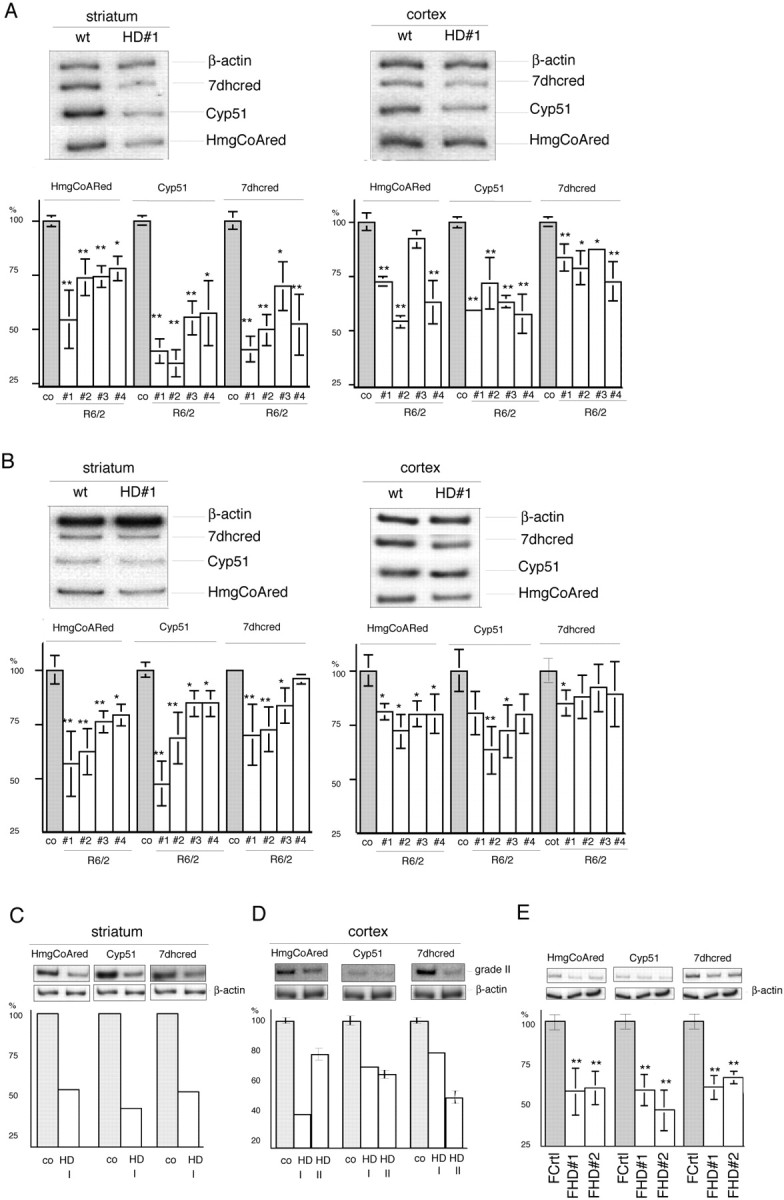
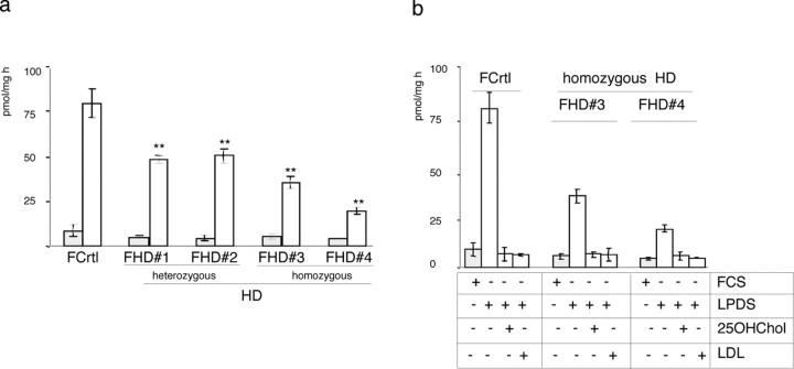
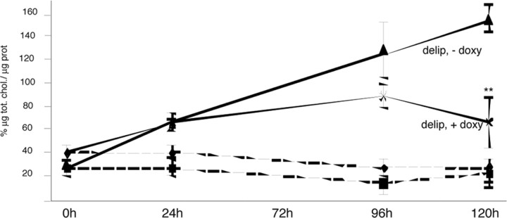
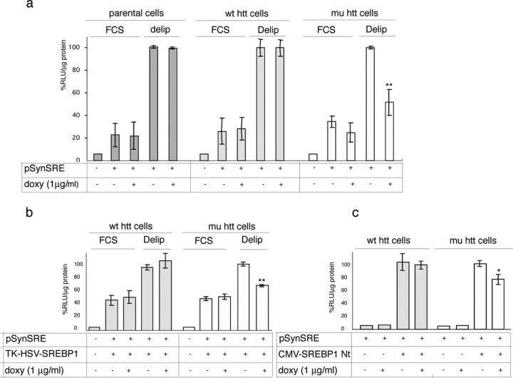
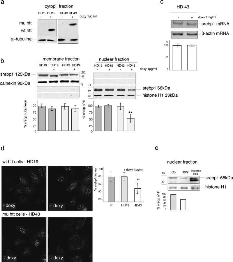
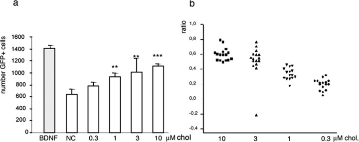
The expansion of a polyglutamine tract in the ubiquitously expressed huntingtin protein causes Huntington's disease (HD), a dominantly inherited neurodegenerative disease. We show that the activity of the cholesterol biosynthetic pathway is altered in HD. In particular, the transcription of key genes of the cholesterol biosynthetic pathway is severely affected in vivo in brain tissue from HD mice and in human postmortem striatal and cortical tissue; this molecular dysfunction is biologically relevant because cholesterol biosynthesis is reduced in cultured human HD cells, and total cholesterol mass is significantly decreased in the CNS of HD mice and in brain-derived ST14A cells in which the expression of mutant huntingtin has been turned on. The transcription of the genes of the cholesterol biosynthetic pathway is regulated via the activity of sterol regulatory element-binding proteins (SREBPs), and we found an approximately 50% reduction in the amount of the active nuclear form of SREBP in HD cells and mouse brain tissue. As a consequence, mutant huntingtin reduces the transactivation of an SRE-luciferase construct even under conditions of SREBP overexpression or in the presence of an exogenous N-terminal active form of SREBP. Finally, the addition of exogenous cholesterol to striatal neurons expressing mutant huntingtin prevents their death in a dose-dependent manner. We conclude that the cholesterol biosynthetic pathway is impaired in HD cells, mice, and human subjects, and that the search for HD therapies should also consider cholesterol levels as both a potential target and disease biomarker.


Figures

Figure 1.
Figure 1.
Reduced HMGCoAred, Cyp51, and 7dhcred gene transcription in mouse and human HD brain and peripheral cells. A, B, Semiquantitative radioactive RT-PCR analyses of striatum and cortex from individual symptomatic 12-week-old (A) and early symptomatic 6-week-old (B) R6/2 transgenic mice compared with the mean values ± SD of three age-matched controls (co). The tissue from each transgenic mouse was investigated by means of six independent RT-PCR analyses. wt, Wild type. C, Human postmortem striatum from grade I HD (HD I). D, Human postmortem cortical tissue from grade I (HD I) and grade II (HD II) versus control. E, Normal (FCrtl) and HD cultured human fibroblasts (FHD). Because of limitations in the available RNA, the grade I striatum and cortex data relate to a single RT-PCR, repeated twice; the data from the grade II samples were obtained by repeating the RT-PCR three times. The graphs show the quantitative analyses of HMGCoAred, Cyp51, and 7dhcred mRNA levels. The peak densitometric area was normalized over the peak densitometric area of the β-actin band. The data are expressed as percentages of control values. *p < 0.05 and **p < 0.01 versus wild-type littermates (co) (A, B), controls (C, D), or normal cultured fibroblasts (FCrtl) (E) (ANOVA). Error bars represent SEM.
Figure 2.
Figure 2.
Modulation of cholesterol biosynthesis in HD cells. a, The activity of the mevalonate pathway was evaluated by incorporating radioactive (14C) acetate into the cellular sterols of cells exposed to medium containing 10% FCS or 10% LPDS. No differences in cholesterol biosynthesis were observed between normal cultured fibroblasts (FCrtl) and HD fibroblasts (FHD) in serum containing medium (dark gray columns). When the cells were exposed to LPDS (light gray columns), cholesterol biosynthesis was significantly reduced in heterozygous and homozygous HD fibroblasts compared with control. **p < 0.01 versus FCrtl (ANOVA). b, Effect of 25-OH-cholesterol (25OHChol) and low-density lipoprotein (LDL) on cholesterol biosynthesis in FCrtl and HD fibroblasts exposed to LPDS. The basal level of cholesterol biosynthesis was restored by the addition of 25OHChol or LDL in both normal and HD cells. The results are normalized to protein content. Mean values ± SD of triplicate experiments. Error bars represent SEM.
Figure 3.
Figure 3.
Total cholesterol (tot. chol.) content in inducible HD cells during exposure to delipidated medium (delip). Total cholesterol levels remained unchanged in induced and uninduced cells cultured in serum containing medium [FCS, -doxy (doxycycline); FCS, +doxy; shaded lines]. Exposure to delipidated medium progressively decreased total cholesterol in the cells expressing mutant huntingtin (delip, +doxy) compared with the same cells under uninduced conditions (delip, -doxy). The results are normalized to protein (prot) content. Mean values ± SD of triplicate experiments. **p < 0.01 versus mutant huntingtin cells grown in the absence of doxy (delip, -doxy; ANOVA). Error bars represent SEM.
Figure 4.
Figure 4.
Impaired SRE activity in HD cells. a, The HMGCoA synthase SRE promoter construct (PSynSRE) was transfected into parental, wild-type huntingtin (wt htt), or mutant huntingtin (mu htt) inducible cells, after which doxycycline (doxy) was added for 24 h and was followed by 16 h of exposure to FCS containing medium or lipid-deprived conditions (delip). Parental cells ± doxy were used as controls. b, The cells were cotransfected with PSynSRE and a construct encoding HSV epitope-tagged human SREBP1a driven by the HSV TK promoter and exposed to complete medium (with FCS) or delipidated conditions. c, Cotransfection of pSynSRE and a construct encoding the N-terminal active form of SREBP1a (CMV-SREBP1a Nt). In this experimental paradigm, 1 μg/ml 25-OH-cholesterol and 10 μg/ml cholesterol (suppressed conditions) were added to the complete medium to suppress the activation of endogenous SREBPs. In all of the luciferase assays, relative light units (RLU) were normalized to protein content, and the data were expressed as the percentages of RLU per microgram of protein from the same cells in the absence of doxy, under delipidated (a, b) or suppressed (c) conditions. Data are mean values of triplicate experiments, one of which is shown. **p < 0.01 versus each cell type grown in the absence of doxy (ANOVA). Error bars represent SEM.
Figure 5.
Figure 5.
Impaired translocation of endogenous SREBP in HD models. a, b, Western blot analyses of endogenous SREBP1 in nuclear and membrane extracts from parental, wild-type huntingtin (wt htt) (HD19), and mutant huntingtin (mu htt) (HD43) inducible cells in the absence or presence of the inducer [1 μg/ml doxycycline (doxy)] in delipidated growth medium. HD43 cells (Q105; mutant clone) and HD19 cells (Q26; normal clone), respectively, overexpress the N-548aa fragment of mutant or wild-type huntingtin. a, Induction of wild-type or mutant huntingtin in the cells after 40 h exposure to doxy. b, Localization of the 125 kDa inactive membrane form and the 68 kDa active nuclear form of SREBP1 in the absence or presence of wild-type or mutant huntingtin, after 16 h in delipidated medium. Calnexin and histone H1 were, respectively, used as loading controls in the membrane and nuclear fractions. The graphs show the quantitative densitometric analyses of the inactive and active forms of SREBP1 compared with calnexin and histone H1. The data are expressed as percentages of the wild-type or mutant cells without doxy. c, Semiquantitative radioactive RT-PCR analysis of mRNA levels of SREBP1 in HD43 after incubation in delipidated growth medium. There were no differences in SREBP1 mRNA levels in the absence or presence of mutant huntingtin. d, Immunocytochemistry for SREBP1 in HD19 and HD43 clones under the same conditions as those described above. The graph shows the mean percentage of nuclear immunoreactivity over the total cell number obtained by counting four microscope fields (10× magnification) from three replicates per experiment. The parental (P) cells (± doxy) behaved like the wild-type huntingtin cells (data not shown). a-d, *p < 0.05 and ** p < 0.01 versus each cell type grown in the absence of doxy (ANOVA). e, Localization of the 68 kDa-active form of SREBP1 in nuclear preparations from HD and control mice. Histone H1 was used as the loading control for the nuclear fraction. The graph shows the quantitative densitometric analysis of the nuclear form of SREBP1 compared with histone H1. The Western blot represents one of five experiments that gave similar results. The data are expressed as percentages of controls (Co); the SD in the graph was obtained by including all of the experiments. **p < 0.01 versus control tissues (ANOVA). Error bars represent SEM.
Figure 6.
Figure 6.
Cholesterol rescues the cell death induced by the transfection of mutant huntingtin in primary neurons. a, Primary rat striatal neurons were prepared and electroporated with plasmids encoding Htt480.68 and GFP and then plated into 96-well microplates. The expression of Htt480.68 and GFP was investigated by means of Western blotting using 1HU-4C8 antibody (Euromedex) for huntingtin and anti-GFP antibody (clone 7.1 and 13.1; Roche Diagnostics) (supplemental Fig. 4, available at www.jneurosci.org as supplemental material). The quantities were normalized to those of β-tubulin revealed using anti-β-tubulin antibody (clone DM1A; Sigma). Eight wells per plate were untreated [negative control (NC)] or treated with 5 ng/ml BDNF or increasing concentrations of cholesterol (chol) dissolved in DMSO. BDNF was used as a positive control. All of the wells received the same final concentration of DMSO (0.5%). Six days after plating, the GFP-positive cells were counted using a fluorescence imaging plate reader (Trophos Flash Cytometer). One of two experiments is shown. b, The combined data from two experiments are expressed as ratios relating to the effect of BDNF. Bonferroni's post hoc analyses revealed significant differences between each tested concentration and the negative control (except for 0.3 μm, as shown in the graph), as well as between each tested concentration and the positive controls. Moreover, among the tested concentrations, significance was reached between 10 and 1 μm, 10 and 0.3 μm, and 3 and 0.3 μm, thus showing the progressive effect of cholesterol dosing. ANOVA also revealed significant differences between treatments (F(5,90) = 34.77; p < 0.0001) and no differences within each treatment group (F(7,40) = 0.048, p = 1 for experiment 1, and F(7,40) = 0.17, p = 0.99 for experiment 2). Error bars represent SEM.

References

    1. Barres BA, Smith SJ (2001) Cholesterol—making or breaking the synapse. Science 294: 1296-1297. - PubMed
    1. Battaile KP, Steiner RD (2000) Smith-Lemli-Opitz syndrome: the first mal-formation syndrome associated with defective cholesterol synthesis. Mol Genet Metab 71: 154-162. - PubMed
    1. Bennett MK, Toth JI, Osborne TF (2004) Selective association of sterol regulatory element-binding protein isoforms with target promoters in vivo. J Biol Chem 279: 37360-37367. - PubMed
    1. Brown AJ, Sun L, Feramisco JD, Brown MS, Goldstein JL (2002) Cholesterol addition to ER membranes alters conformation of SCAP, the SREBP escort protein that regulates cholesterol metabolism. Mol Cell 10: 237-245. - PubMed
    1. Brown MS, Goldstein JL (1997) The SREBP pathway: regulation of cholesterol metabolism by proteolysis of a membrane-bound transcription factor. Cell 89: 331-340. - PubMed

Publication types

MeSH terms

Substances