Comparative Study
. 2005 Oct 26;25(43):9940-8.
doi: 10.1523/JNEUROSCI.3467-05.2005.

Vascular development of the brain requires beta8 integrin expression in the neuroepithelium

Affiliations
Comparative Study

Vascular development of the brain requires beta8 integrin expression in the neuroepithelium

John M Proctor et al. J Neurosci. .

Abstract

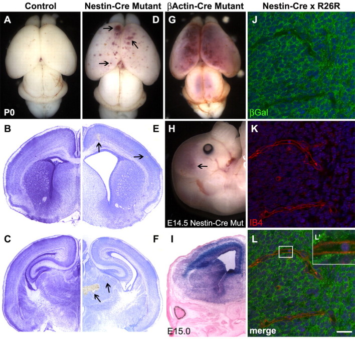
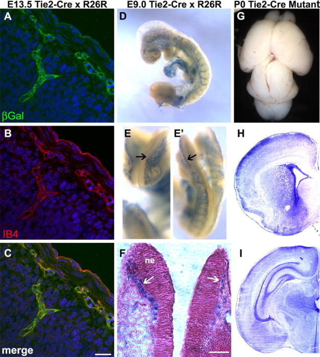
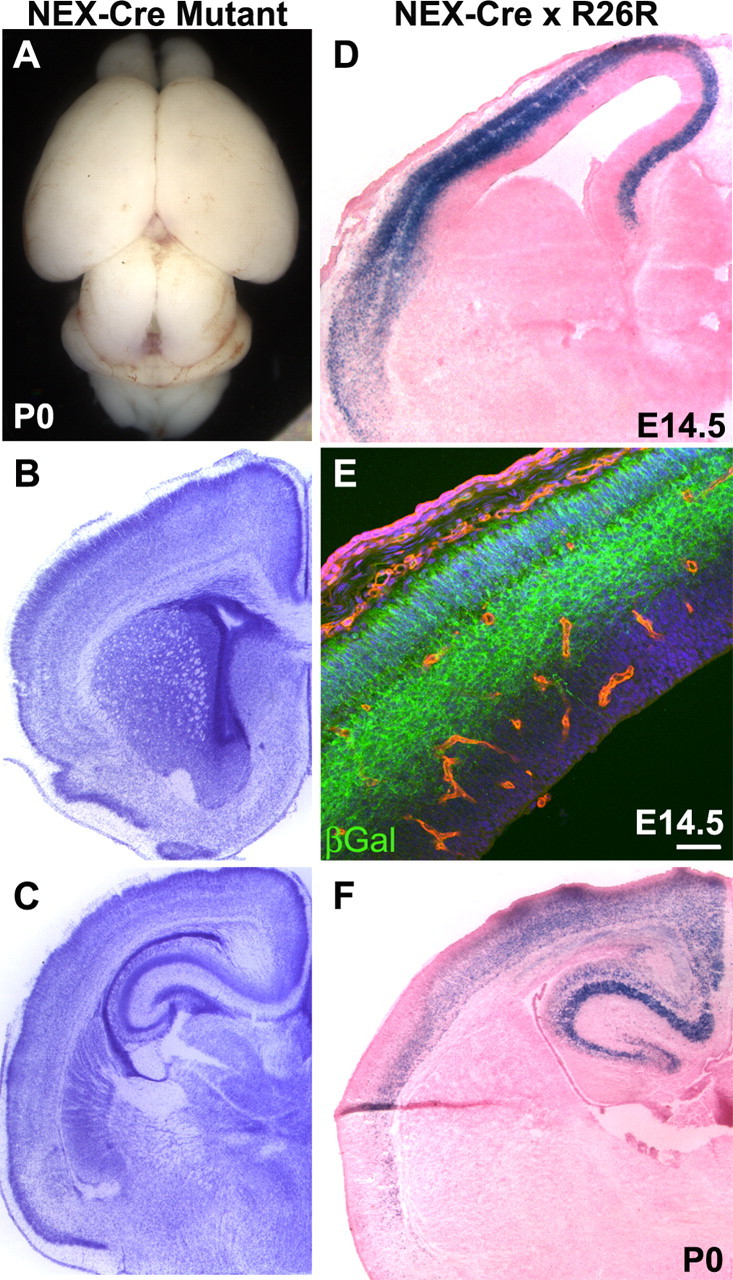
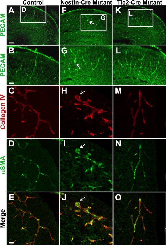
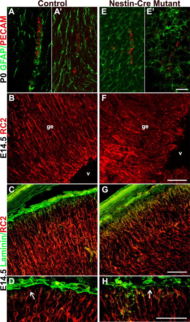
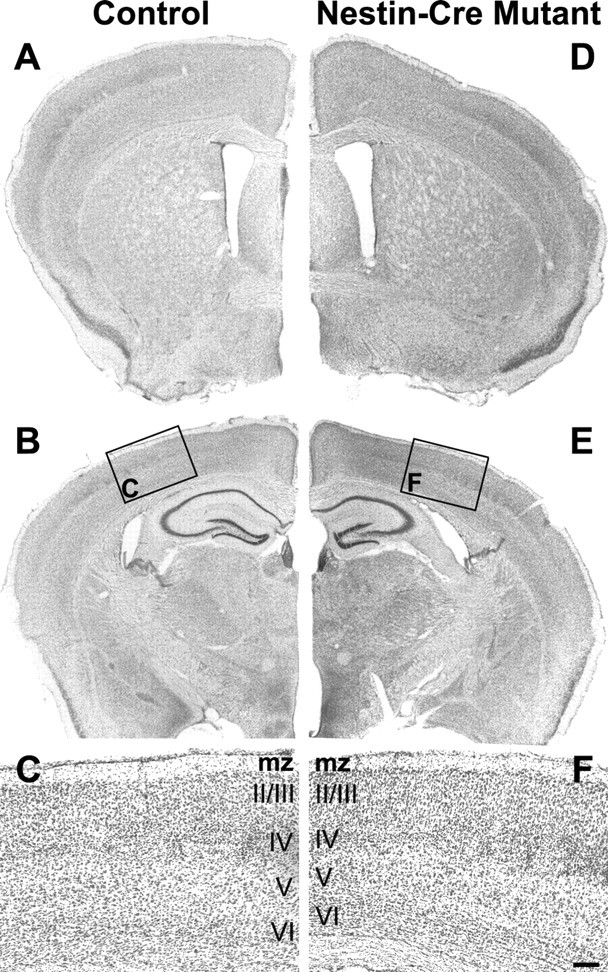
We showed previously that loss of the integrin beta8 subunit, which forms alphavbeta8 heterodimers, results in abnormal vascular development in the yolk sac, placenta, and brain. Animals lacking the integrin beta8 (itgbeta8) gene die either at midgestation, because of insufficient vascularization of the placenta and yolk sac, or shortly after birth with severe intracerebral hemorrhage. To specifically focus on the role of integrins containing the beta8 subunit in the brain, and to avoid early lethalities, we used a targeted deletion strategy to delete itgbeta8 only from cell types within the brain. Ablating itgbeta8 from vascular endothelial cells or from migrating neurons did not result in cerebral hemorrhage. Targeted deletion of itgbeta8 from the neuroepithelium, however, resulted in bilateral hemorrhage at postnatal day 0, although the phenotype was less severe than in itgbeta8-null animals. Newborn mice lacking itgbeta8 from the neuroepithelium had hemorrhages in the cortex, ganglionic eminence, and thalamus, as well as abnormal vascular morphogenesis, and disorganized glia. Interestingly, adult mice lacking itgbeta8 from cells derived from the neuroepithelium did not show signs of hemorrhage. We propose that defective association between vascular endothelial cells and glia lacking itgbeta8 is responsible for the leaky vasculature seen during development but that an unidentified compensatory mechanism repairs the vasculature after birth.


Figures

Figure 1.
Figure 1.
Phenotypic defects observed after conditional deletion of itgβ8 from the neuroepithelium using nestin-cre. A, Wild-type brain from P0 neonate. B, C, Nissl-stained sections of a control brain. D, P0 brain lacking itgβ8 because of conditional deletion mediated by neuroepithelial-specific nestin-cre. Note the blood produced from hemorrhages throughout the dorsal and rostral cortices (arrowheads). E, F, Nissl-stained sections of a nestin-cre mutant brain. Notice the hemorrhages (arrowheads) throughout the cortex (E) in addition to the massive hemorrhage in the thalamus (F). G, P0 mutant animal generated using the β-actin-cre line. Mutants generated with this cre line display phenotypes not obviously different from the null animal. An avas-cularized yolk sac and placenta is seen in the majority of embryos that die by E11.5, and severe hemorrhage is seen in the brain of embryos that survive to birth. H, E14.5 brain lacking itgβ8 from the neuroepithelium because of conditional deletion mediated by neuroepithelial-specific nestin-cre. Note the blood produced from hemorrhages throughout the dorsal and rostral cortices (arrowhead). I, LacZ-stained section from an E15 nestin-cre-positive animal crossed to the R26R reporter strain. Note that the neuroepithelium is almost entirely recombined. J-L′, β-galactosidase expression analysis of E15 embryo cortices (one-half day after hemorrhage was observed in nestin-cre mutants) obtained from a cross of a nestin-cre-positive animal and the R26R reporter line. Note the strong expression of β-galactosidase (green) in cells of the neuroepithelium but the lack of β-galactosidase immunolabeling of the endothelial cells labeled with isolectin B4 (red) (L′). Cell nuclei were counterstained with TO-PRO-3 (blue). βGal, β-Galactosidase; Mut, mutant; IB4, isolectin B4. Scale bar: L, 20 μm.
Figure 2.
Figure 2.
Conditional deletion of itgβ8 from vascular endothelial cells using tie2-cre does not result in cerebral hemorrhage. A-C, β-Galactosidase (βGal) expression analysis of E13.5 embryo cortices obtained from a cross between a tie2-cre-positive animal and the R26R reporter line. Note that β-galactosidase (green) expression is very strong and perfectly overlaps with the isolectinB4 (IB4) (red) (B, C) immunolabeling of endothelial cells. Cell nuclei were counterstained with TO-PRO-3 (blue). D-F, LacZ reporter analysis of E9 embryos obtained from a cross between a tie2-cre-positive animal and the R26R reporter line. Notice that cre-mediated recombination is very strong in the head vasculature before invasion of the neuroepithelium (E, E′, arrows). F, Coronal slice of embryo from E, counterstained with Nuclear Fast Red. Notice that LacZ-positive vessels (blue) have not yet invaded the neuroepithelium (ne) at 9.0 d postconception. G, P0 brain lacking itgβ8 in vascular endothelial cells because of conditional deletion mediated by tie2-cre. H, I, Nissl-stained sections of a tie2-cre mutant P0 brain. Note the absence of hemorrhage. Scale bars: C, 20 μm; F, 40 μm.
Figure 3.
Figure 3.
Conditional deletion of itgβ8 from postmitotic neurons using nex-cre does not result in cerebral hemorrhage. A, P0 brain lacking itgβ8 in postmitotic neurons because of conditional deletion mediated by nex-cre. B, C, Nissl-stained sections of a nex-cre mutant P0 brain. D, F, LacZ-stained sections from an E14.5 embryo cortex and P0 cortex obtained from a cross between a nex-cre-positive animal and the R26R reporter line. Cre-mediated recombination is very strong in the future cortical plate by E14.5 when hemorrhage is observed in the nestin-cre mutant and remains strong after birth. E, β-Galactosidase (βGal) expression analysis of E14.5 embryo cortex. Note that βGal (green) immunolabeling is limited to cells within the future cortical plate. Endothelial cells were immunolabeled with isolectin B4 (red) and were not recombined by this cre line. Cell nuclei were counterstained with TO-PRO-3 (blue). Scale bar: E, 50 μm.
Figure 4.
Figure 4.
Conditional deletion of itgβ8 in the neuroepithelium leads to endothelial cell irregularities in the P0 neonatal cortex. Anti-PECAM immunolabeling of P0 coronal sections of the forebrain to visualize vascular endothelial cells is shown. A, B, Control. F, G, Nestin-cre-targeted mutant. K, L, Tie2-cre-targeted mutant. Notice the bulbous organization of endothelial cell clusters in the cortex (arrows) of the nestin-cre-targeted mutant (F, G). Anti-collagen IV immunolabeling of P0 coronal sections of the neuroepithelium to visualize the basal lamina surrounding the vasculature is shown. C, E, Control. H, J, Nestin-cre-targeted mutant. M, O, Tie2-cre-targeted mutant. Notice the intact basement membrane in the area of an endothelial cell cluster (arrowhead) in the nestin-cre-targeted mutant (H, J). Anti-α-smooth muscle actin (αSMA) immunolabeling of pericytes in P0 coronal sections of the neuroepithelium. D, E, Control. I, J, Nestin-cre mutant. N, O, Tie2-cre mutant. Note the normal recruitment of pericytes to the vasculature in both mutants. Scale bars: A, 80 μm; B, E, 40 μm.
Figure 5.
Figure 5.
Itgβ8 nestin-cre mutants have abnormally organized cortical glia. Anti-PECAM/GFAP immunostaining of endothelial cells and glia, respectively, in P0 coronal sections of the dorsal forebrain is shown. A, A′, Control. E, E′, Nestin-cre mutant. Notice the disorganization of the astroglia and the lack of alignment between endothelial cells and the astroglial processes (E, E′). Anti-RC2 immunostaining of radial glia in E14.5 coronal sections of the ganglionic eminence is shown. B, Control. F, Nestin-cre-targeted mutant. Radial glia in F are badly disorganized, most likely because of hemorrhage within the ganglionic eminence (ge) near the lateral ventricles (v). Anti-RC2 and anti-laminin immunolabeling of E14.5 coronal sections demonstrating radial glia cell morphology in cortices of a control animal (C, D) and a nestin-cre mutant (G, H) is shown. Attachment of the radial glia to the pial surface in the nestin-cre mutant does not appear altered (H, arrow), and glial processes display normal organization. Scalebars: E′, 20 μm; F-H, 50 μm.
Figure 6.
Figure 6.
Adult itgβ8 nestin-cre mutants show no sign of brain hemorrhage. A, Six-week-old control. D, Six-week-old nestin-cre-targeted mutant coronal sections at the level of the lateral ventricles in the forebrain stained with Nissl. B, Control. E, Nestin-cre mutant coronal sections at the level of the hippocampus stained with Nissl. Note the lack of hemorrhage or other obvious defect in the cortex or thalamus. C, F, High magnification of cortical sections boxed in B and E. All cortical layers are present and in the correct order in the mutant. mz, Marginal zone. Scale bar: F, 100 μm.

References

    1. Abbott NJ, Romero IA (1996) Transporting therapeutics across the blood-brain barrier. Mol Med Today 2: 106-113. - PubMed
    1. Bader BL, Rayburn H, Crowley D, Hynes RO (1998) Extensive vasculogenesis, angiogenesis, and organogenesis precede lethality in mice lacking all alpha v integrins. Cell 95: 507-519. - PubMed
    1. Beggs HE, Schahin-Reed D, Zang K, Goebbels S, Nave KA, Gorski J, Jones KR, Sretavan D, Reichardt LF (2003) FAK deficiency in cells contributing to the basal lamina results in cortical abnormalities resembling congenital muscular dystrophies. Neuron 40: 501-514. - PMC - PubMed
    1. Breier G, Albrecht U, Sterrer S, Risau W (1992) Expression of vascular endothelial growth factor during embryonic angiogenesis and endothelial cell differentiation. Development 114: 521-532. - PubMed
    1. Brockschnieder D, Lappe-Siefke C, Goebbels S, Boesl MR, Nave KA, Riethmacher D (2004) Cell depletion due to diphtheria toxin fragment A after Cre-mediated recombination. Mol Cell Biol 24: 7636-7642. - PMC - PubMed

Publication types

MeSH terms