Comparative Study
. 2005 Oct 26;25(43):10029-40.
doi: 10.1523/JNEUROSCI.2652-05.2005.

Partial mitochondrial inhibition causes striatal dopamine release suppression and medium spiny neuron depolarization via H2O2 elevation, not ATP depletion

Affiliations
Comparative Study

Partial mitochondrial inhibition causes striatal dopamine release suppression and medium spiny neuron depolarization via H2O2 elevation, not ATP depletion

Li Bao et al. J Neurosci. .

Abstract

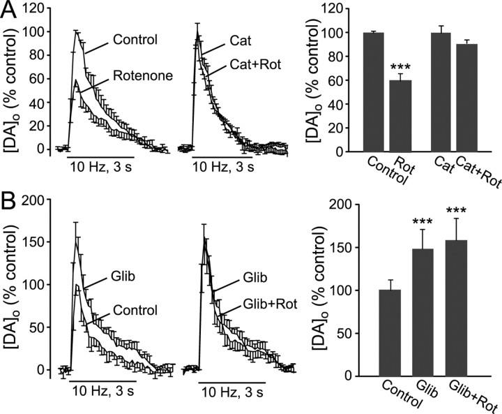
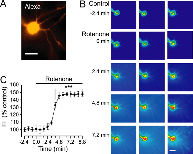
Mitochondrial dysfunction is a potential causal factor in Parkinson's disease. We show here that acute exposure to the mitochondrial complex I inhibitor rotenone (30-100 nM; 30 min) causes concentration-dependent suppression of single-pulse evoked dopamine (DA) release monitored in real time with carbon-fiber microelectrodes in guinea pig striatal slices, with no effect on DA content. Suppression of DA release was prevented by the sulfonylurea glibenclamide, implicating ATP-sensitive K+ (KATP) channels; however, tissue ATP was unaltered. Because KATP channels can be activated by hydrogen peroxide (H2O2), as well as by low ATP, we examined the involvement of rotenone-enhanced H2O2 generation. Confirming an essential role for H2O2, the inhibition of DA release by rotenone was prevented by catalase, a peroxide-scavenging enzyme. Striatal H2O2 generation during rotenone exposure was examined in individual medium spiny neurons using fluorescence imaging with dichlorofluorescein (DCF). An increase in intracellular H2O2 levels followed a similar time course to that of DA release suppression and was accompanied by cell membrane depolarization, decreased input resistance, and increased excitability. Extracellular catalase markedly attenuated the increase in DCF fluorescence and prevented rotenone-induced effects on membrane properties; membrane changes were also largely prevented by flufenamic acid, a blocker of transient receptor potential (TRP) channels. Thus, partial mitochondrial inhibition can cause functional DA denervation via H2O2 and KATP channels, without DA or ATP depletion. Furthermore, amplified H2O2 levels and TRP channel activation in striatal spiny neurons indicate potential sources of damage in these cells. Overall, these novel factors could contribute to parkinsonian motor deficits and neuronal degeneration caused by mitochondrial dysfunction.


Figures

Figure 1.
Figure 1.
Rotenone causes suppression of axonal DA release in dorsal striatum. A, Representative DA release records elicited by a single stimulus pulse under control conditions and after 30 min exposure to rotenone (50 nm) at the same recording site. Inset, Cyclic voltammograms taken at the maximum [DA]o for each record; dopamine was identified by characteristic oxidation (Ox) and reduction (Red) peak potentials (typically +600 and -200 mV vs Ag/AgCl). B, Time course of rotenone-induced suppression of single-pulse evoked [DA]o elicited at 2.5 min intervals throughout rotenone exposure (n = 10; *p < 0.05; **p < 0.01 rotenone vs control; ANOVA). C, Concentration dependence of rotenone-induced suppression of single-pulse evoked DA release. Release suppression induced by rotenone at 30 nm (n = 6), 50 nm (n = 10), or 100 nm (n = 6) differed significantly at the time points indicated (n = 6; **p < 0.01). Error bars indicate SE.
Figure 2.
Figure 2.
Rotenone-induced suppression of DA release requires KATP channel opening. A, Average DA release records after single-pulse stimulation elicited at 5 min intervals under control conditions and after 30 min exposure to rotenone (50 nm; n = 7) compared with release in the presence of glibenclamide (Glib) (3 μm) and glibenclamide plus rotenone (n = 5). Data are normalized, with maximum evoked [DA]o under control conditions for each slice taken as 100%. B, Rotenone (Rot) causes a decrease in evoked [DA]o (***p < 0.001 vs control; n = 7). Glibenclamide had no effect on single-pulse evoked DA release; however, this KATP channel blocker completely prevented the usual suppression of DA release in the presence of rotenone (n = 5; p > 0.05 vs glibenclamide alone). C, Average single-pulse evoked [DA]o under control conditions, in the presence of tolbutamide (Tolb) (200 μm), and after 30 min exposure to rotenone (50 nm) in the presence of tolbutamide (n = 3). D, Tolbutamide had no effect on single-pulse evoked DA release; however, it completely prevented DA release suppression by rotenone (n = 3; p > 0.05 vs tolbutamide alone). Error bars indicate SE.
Figure 3.
Figure 3.
Acute rotenone exposure does not alter striatal ATP content. Average ATP content in dorsal striatum from striatal slices exposed to rotenone (50 nm) for 5, 10, 20, or 30 min. Data are given as percentage control (% control), with the ATP content of paired slices incubated for the same duration in ACSF alone taken as 100%. Tissue ATP content did not differ between rotenone-exposed and control slices at any time point (p > 0.05 vs paired control; n = 10-12). Error bars indicate SE.
Figure 4.
Figure 4.
Evoked DA release is unaltered by a drop in ATP caused by low glucose. A, ATP content in dorsal striatum from slices maintained in the recording chamber in normal ACSF with 10 mm glucose (Control) and/or in modified ACSF with low glucose (1 mm) for 30 min, given as percentage control (% control). Incubation in low glucose caused a significant decrease in striatal ATP content (***p < 0.001 vs paired control; n = 13). B, Average DA concentration-time profiles in dorsal striatum after single-pulse stimulation elicited at 5 min intervals in normal ACSF, after 30 min in ACSF with 1 mm glucose, and after 30 min washout with normal ACSF (Wash) (n = 5). C, Maximum evoked [DA]o did not differ between 1 mm and 10 mm glucose-containing ACSF (n = 5; p > 0.05, 1 mm vs either control or wash). Error bars indicate SE.
Figure 5.
Figure 5.
Rotenone-induced suppression of DA release requires H2O2. A, Average DA release records elicited by single-pulse stimulation under control conditions (Control) and after 30 min exposure to rotenone (Rot) (50 nm) compared with the effect of rotenone on single-pulse release in the presence of catalase (Cat) (500 U/ml). B, Under control conditions (ACSF alone or ACSF plus heat-inactivated catalase), rotenone caused a significant decrease in peak evoked [DA]o (***p < 0.001, rotenone vs control; n = 9); however, this effect of rotenone was prevented in the continued presence of catalase (p > 0.05 vs same-site control in catalase alone; n = 5). Error bars indicate SE.
Figure 6.
Figure 6.
Enhanced suppression of DA release by rotenone with pulse-train stimulation also requires H2O2 and KATP channel opening. A, Average DA release records elicited by 30 pulses (10 Hz) under control conditions (Control) and after 30 min exposure to rotenone (Rot) (50 nm) compared with the effect of rotenone on pulse-train evoked [DA]o in the presence of catalase (Cat) (500 U/ml). Under control conditions (ACSF alone or ACSF plus heat-inactivated catalase), rotenone caused a significant decrease in peak evoked [DA]o (***p < 0.001 rotenone vs control; n = 7); however, this effect of rotenone was prevented in the continued presence of catalase (p > 0.05 vs same-site control in catalase alone; n = 5). B, Average pulse-train evoked DA release records under control conditions, in the presence of glibenclamide (Glib) (3 μm), and after rotenone in glibenclamide. Glibenclamide caused an increase in pulse-train evoked [DA]o (***p < 0.001 vs control; n = 5) and prevented rotenone-induced suppression of DA release (p > 0.05 vs glibenclamide alone; ***p < 0.001 vs control). Error bars indicate SE.
Figure 7.
Figure 7.
Rotenone increases H2O2 generation in striatal medium spiny neurons. A, Medium spiny neuron visualized with Alexa Red during physiological recording in vitro. B, DCF fluorescence in the cell in A under control conditions and during exposure to rotenone (50 nm). Scale bars: A, B, 20 μm. C, Average time course and amplitude of H2O2 generation [DCF fluorescence intensity (FI)] during rotenone exposure (n = 13; ***p < 0.001 rotenone vs basal FI; ANOVA). Error bars indicate SE.
Figure 8.
Figure 8.
Rotenone alters membrane properties of striatal medium spiny neurons. A-D, Representative current-clamp records during a series of hyperpolarizing and depolarizing current pulses (0.04 nA; 10-12 steps) in medium spiny neurons in striatal slices in vitro. A, B, Current-clamp records from the cell shown in Figure 7, A and B; the pattern of membrane responses under control conditions (A) confirmed the identity of the cell as a medium spiny neuron. Rotenone exposure (50 nm; ≤30 min) increased cell excitability (n = 13) (B), as described in Results. C, D, Current-clamp records from a different medium spiny neuron in the presence of catalase (500 U/ml) and in catalase plus rotenone. In the presence of catalase, rotenone (50 nm; ≤30 min) had no effect on membrane properties in medium spiny neurons (n = 3). The usual increase in cell excitability with rotenone was seen in heat-inactivated catalase (n = 3); data from heat-inactivated catalase were included in control data averages.
Figure 9.
Figure 9.
Acute rotenone exposure alters membrane properties of medium spiny neurons. A, Representative current-clamp record showing membrane depolarization in a striatal medium spiny neuron during application of 50 nm rotenone; resting potential in this cell was -78.4 mV (dashed line). Each downward deflection represents the voltage drop produced by pulsed current injection (-0.1 nA; 1 s) used to monitor input resistance during the experiments; the decreasing amplitude of these deflections indicated that a decrease in input resistance accompanied rotenone application. B, Time course of rotenone-induced membrane potential (Memb pot'l) changes in striatal spiny neurons during exposure to 50 nm rotenone (n = 10; *p < 0.05; **p < 0.01 rotenone vs control; ANOVA). C, Time course of rotenone-induced changes in input resistance (R) for the cells included in B (n = 10; **p < 0.01 rotenone vs control; ANOVA). D, Effect of blocking TRP channels with FFA (10 μm) or blocking KATP channels with glibenclamide (Glib) (3 μm) on rotenone-induced changes in membrane in striatal spiny neurons. Marked suppression of rotenone-induced changes in the presence of FFA implicates activation of TRP channels as the underlying cause of rotenone-induced membrane depolarization in medium spiny neurons (n = 5; ***p < 0.001 vs same time point in 50 nm rotenone alone). Enhanced depolarization in the presence of glibenclamide indicates that activation of KATP channels during rotenone exposure normally opposes TRP-channel-mediated depolarization (n = 5; ***p < 0.001 vs same time point in 50 nm rotenone alone). E, Effect of FFA (n = 5) or glibenclamide (n = 5) on rotenone-induced changes in input resistance for the cells included in D; both TRP and KATP channels contribute to decreased membrane resistance (increased conductance), in the presence of rotenone (***p < 0.001 vs same time point in 50 nm rotenone alone). Error bars indicate SE.

Similar articles

Cited by

References

    1. Alam M, Schmidt WJ (2004) l-DOPA reverses the hypokinetic behaviour and rigidity in rotenone-treated rats. Behav Brain Res 153: 439-446. - PubMed
    1. Ashcroft FM, Gribble FM (1998) Correlating structure and function in ATP-sensitive K+ channels. Trends Neurosci 21: 288-294. - PubMed
    1. Avshalumov MV, Rice ME (2003) Activation of ATP-sensitive K+ (KATP) channels by H2O2 underlies glutamate-dependent inhibition of striatal dopamine release. Proc Natl Acad Sci USA 100: 11729-11734. - PMC - PubMed
    1. Avshalumov MV, Chen BT, Marshall SP, Peña DM, Rice ME (2003) Glutamate-dependent inhibition of dopamine release in striatum is mediated by a new diffusible messenger, H2O2 J Neurosci 23: 2744-2750. - PMC - PubMed
    1. Avshalumov MV, Chen BT, Koós T, Tepper JM, Rice ME (2005) Endogenous hydrogen peroxide regulates the excitability of midbrain dopamine neurons via ATP-sensitive channels. J Neurosci 25: 4222-4231. - PMC - PubMed

Publication types

MeSH terms

LinkOut - more resources