. 2005 Nov 14;93(10):1157-67.
doi: 10.1038/sj.bjc.6602831.

Restoration of TGF-beta signalling reduces tumorigenicity in human lung cancer cells

Affiliations

Restoration of TGF-beta signalling reduces tumorigenicity in human lung cancer cells

G Anumanthan et al. Br J Cancer. .

Abstract

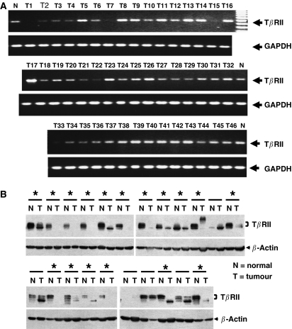
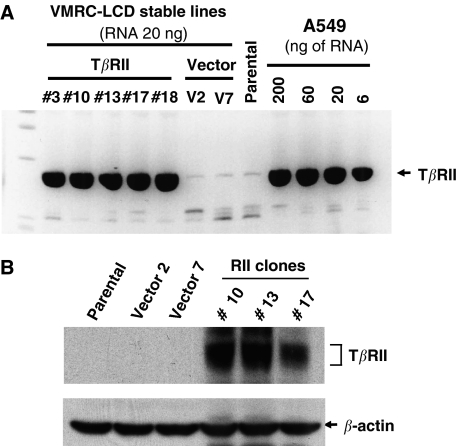
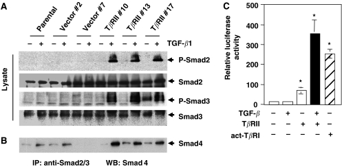
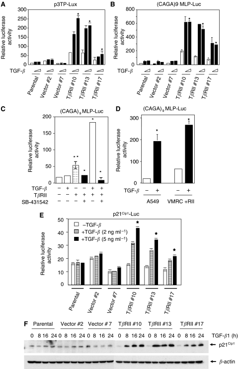
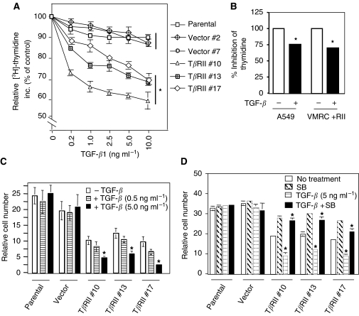
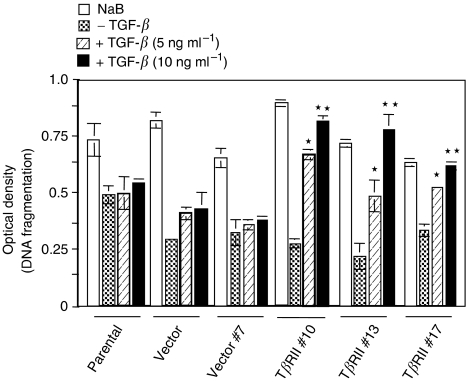
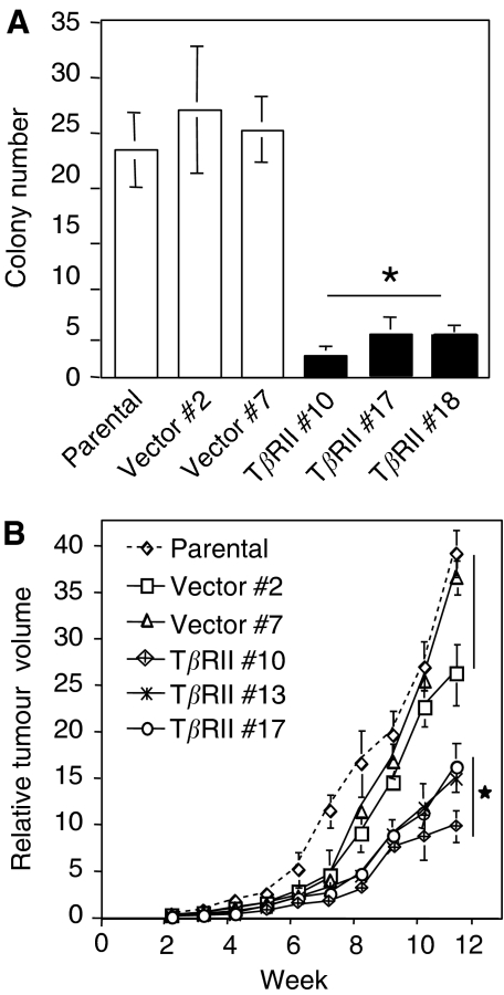
Members of the transforming growth factor-beta (TGF-beta) family regulate a wide range of biological processes including cell proliferation, migration, differentiation, apoptosis, and extracellular matrix deposition. Resistance to TGF-beta-mediated tumour suppressor function in human lung cancer may occur through the loss of type II receptor (TbetaRII) expression. In this study, we investigated the expression pattern of TbetaRII in human lung cancer tissues by RT-PCR and Western blot analyses. We observed downregulation of TbetaRII in 30 out of 46 NSCLC samples (65%) by semiquantitative RT-PCR. Western blot analyses with tumour lysates showed reduced expression of TbetaRII in 77% cases. We also determined the effect of TbetaRII expression in lung adenocarcinoma cell line (VMRC-LCD) that is not responsive to TGF-beta due to lack of TbetaRII expression. Stable expression of TbetaRII in these cells restored TGF-beta-mediated effects including Smad2/3 and Smad4 complex formation, TGF-beta-responsive reporter gene activation, inhibition of cell proliferation and increased apoptosis. Clones expressing TbetaRII showed reduced colony formation in soft-agarose assay and significantly reduced tumorigenicity in athymic nude mice. Therefore, these results suggest that reestablishment of TGF-beta signalling in TbetaRII null cells by stable expression of TbetaRII can reverse malignant behaviour of cells and loss of TbetaRII expression may be involved in lung tumour progression.


Figures

Figure 1
Figure 1
Downregulation of TβRII mRNA in lung tumours as determined by RT–PCR. (A) Total RNA extracted from human lung tumour tissues were reverse transcribed, and the resulting cDNA was analysed by PCR to test TβRII expression. The integrity and equal loading of the RT products was assessed by analysing hGAPDH expression. (B) Western blot analysis of TβRII protein in lung tumour and corresponding normal lung tissues. Equal amount of lysates were resolved in SDS–PAGE and analysed by Western blotting using antibody against TβRII. Equal loading was verified by Western blotting using mouse monoclonal anti-β-actin antibody. Tumours with asterisk showed reduced expression of TβRII.
Figure 2
Figure 2
Stable VMRC-LCD cells expressing TβRII. (A) The VMRC-LCD cells were transfected with TβRII-pcDNA3 or empty pcDNA3 vector (Invitrogen) and selected with G418 for 2 weeks to establish the stable clones. In all, 20 ng of total RNA was used for TβRII stable VMRC-LCD clones, vector clones and parental cells. Quantitative real-time RT–PCR was performed to determine the relative mRNA expression level of each TβRII stable clones. Dilutions of RNA isolated from A549 cells was used as standards for comparison. The stable expression of TβRII mRNA in VMRC-LCD clones were compared with the endogenous TβRII expression in A549 cells. (B) Cell lysates from parental, vector clone, and stable TβRII clones were subjected to immunoblotting with anti-TβRII antibody. Expression of TβRII protein in individual clones is shown. Equal amount of protein loading was verified by immunoblotting the membrane with anti-β-actin antibody.
Figure 3
Figure 3
Stable expression of TβRII in VMRC-LCD cells restores TGF-β-induced phosphorylation of Smad2 and Smad3 and complex formation with Smad4. (A) Parental cells, vector control and stable TβRII clones were preincubated for 2 h in serum-free medium and then treated with TGF-β1 (5 ng ml−1) for 90 min. Cell lysates were subjected to immunoblotting with antiphospho Smad2, anti-Smad2, antiphospho Smad3, anti-Smad3, and anti-Smad4 antibodies. Equal amount of protein loading was tested by immunoblotting the membrane with anti-β-actin antibody. (B) Parental cells, vector control, and stable TβRII clones were treated with TGF-β as above. Equal amount of cell lysates were subjected to immunoprecipitation with anti-Smad2 and anti-Smad3 polyclonal antibodies and the immunoprecipitates were analysed by immunoblotting with anti-Smad4 antibodies. (C) VMRC-LCD parental cells were transiently transfected with (CAGA)9 MLP-Luc, and CMV-β-gal, TβRII or act-TβRI (T204D) expression plasmids. Cells were treated with 5 ng ml−1 TGF-β for 22 h. Luciferase activity was normalised to β-gal activity, and the relative luciferase activity was expressed as the mean±s.d. of triplicate measurements. These experiments were repeated at least three times. *P<0.001 for all groups in a multiple comparison test with Bonferroni adjustments after rank transforming the data.
Figure 4
Figure 4
Restoration of TGF-β-induced transcriptional responses by stable expression of TβRII. Parental, vector control, and TβRII stable clones were transiently cotransfected with CMV-β-Gal, p3TP-Lux (A) or (CAGA)9 MLP-Luc (B) plasmids. At 20 h after transfection, cells were treated with TGF-β1 (2 or 5 ng ml−1) for an additional 22 h in low serum (0.2% FBS)-containing medium. Luciferase activity was normalised to β-gal activity, and the relative luciferase activity was expressed as the mean±s.d. of triplicate measurements. *P<0.0001 for all groups in (A) and (B) in a linear mixed effect model on the log-transformed data. (C) VMRC-LCD parental cells were transfected with CMV-β-Gal, CMV-TβRII, and (CAGA)9 MLP-Luc plasmids. At 20 h after transfection, cells were treated with 5 ng ml−1 TGF-β in the presence or absence of SB-431542 (10 μM) for an additional 22 h in low serum (0.2% FBS)-containing medium. Luciferase activity was normalised to β-gal activity, and the relative luciferase activity was expressed as the mean±s.d. of triplicate measurements. (D) Stable TβRII clone and A549 parental cells were transiently cotransfected with CMV-β-Gal and (CAGA)9 MLP-Luc plasmids. Cells were treated with TGF-β1 (5 ng ml−1) for 22 h as above. Luciferase activity was normalised to β-gal activity, and the relative luciferase activity was expressed as the mean±s.d. of triplicate measurements. **P<0.05, *P<0.005 for all groups in (C) and (D) in a multiple comparison test with Bonferroni adjustments after rank transforming the data. (E) Cells were transiently transfected with CMV-β-gal and p21Cip1-Luc plasmid and treated with 2 or 5 ng ml−1 of TGF-β1 for 22 h. Luciferase activity was normalised to β-gal activity, and the relative luciferase activity was expressed as the mean±s.d. of triplicate measurements. *P<0.001 for all groups in a linear mixed effect model on the log-transformed data. (F) Parental, vector control, and TβRII stable clones were serum starved for 16 h and treated with 5 ng ml−1 of TGF-β1 for different time points. Cell lysates were analysed by Western blotting with anti-p21Cip1 antibody (Santa Cruz Biotechnology). Equal amount of protein loading was verified by Western blotting with anti-β-actin antibody. Each experiment was repeated three times with similar results.
Figure 5
Figure 5
Expression of TβRII induces TGF-β-mediated growth inhibition. Thymidine Incorporation Assay. (A) 30 000 cells well−1 of VMRC-LCD in 24-well plate were treated with increasing doses of TGF-β in presence of 10% FBS-containing medium for 36 h. 4 μCi well−1 [3H]-thymidine (NEN) was added in each well for an additional 4 h. Cells were then fixed, lysed, and the radioactivity incorporated was counted. Radioactivity incorporated without TGF-β treatment is considered as 100%, and the results are expressed as the mean±s.d. for triplicate measurements. (B) Stable TβRII clone and A549 cells were plated as above and treated with TGF-β for 36 h. Cells were processed as above and the radioactivity incorporated was counted. Radioactivity incorporated without TGF-β treatment is considered as 100%, and the results are expressed as the mean±s.d. for triplicate measurements. *P<0.05 for all groups in (A) and (B) were compared by Wilcoxon's test. (C) Cell counting assay. In all, 8 × 10³ cells from parental, vector control, and stable TβRII clones were seeded into each well of 12-well plate and then treated with TGF-β (0.5 or 5 ng ml−1) in 10% FBS-containing medium. Cells were counted after 5 days and plotted. Each data point is expressed as the mean±s.d. of triplicate measurements. Each experiment was repeated three times with similar results. (D) VMRC-LCD parental, vector control, and TβRII stable clones were treated with 5 ng ml−1 TGFβ in the presence or absence of SB-431542 (10 μM) for 5 days. Cells were counted and the cell numbers were plotted. Individual data points are the mean±s.d. of triplicate determinations. *P<0.005 for all groups in (C) and (D) in a linear mixed effect model on the log-transformed data.
Figure 6
Figure 6
Quantitative cell death ELISA. Parental, vector control, and TβRII stable clones were serum starved and treated with either TGF-β1 (5 or 10 ng ml−1) or 1 μM sodium butyrate for 24 h in serum-free media. Cell lysates were analysed by cell death ELISA as described in Materials and methods. Individual data point is a representative of the mean±s.d. of three individual measurements. Each experiment was repeated three times with similar results. *P<0.0008, **P<0.0001 for all groups in a linear mixed effect model on the log-transformed data.
Figure 7
Figure 7
Stable expression of TβRII decreases tumorigenicity. (A) Restoration of TβRII reduces anchorage-independent growth in VMRC-LCD cells. Parental, vector control, and TβRII stable clones were plated in soft agarose and incubated for 2 weeks. Colonies were counted by automated colony counter and the data are representative of the mean±s.d. of three values determined from individual plates. *P<0.005 for all groups was compared to control by Wilcoxon test. (B) Xenograft growth curves of parental, vector control, and TβRII stable clones. Cells (5 × 10⁶) from each pool were subcutaneously injected to the athymic nude mice. Tumours were measured externally on the indicated days in two dimensions using slide calipers. Tumour volume was determined from the equation: V=(L × W2) × 0.5, where L is length and W is width of the tumour. Each data point represents a mean volume±s.e. of six tumours for each group. *P<0.05 for all groups was compared to control by Wilcoxon test.

References

    1. Arteaga CL, Tandon AK, VonHoff DD, Osborne CK (1988) Transforming growth factor beta: potential autocrine growth inhibitor of estrogen receptor-negative human breast cancer cells. Cancer Res 48: 3898–3904 - PubMed
    1. Attisano L, Wrana JL (1998) Mads and Smads in TGF beta signalling. Curr Opin Cell Biol 10: 188–194 - PubMed
    1. Chen CR, Kang Y, Massague J (2001) Defective repression of c-myc in breast cancer cells: A loss at the core of the transforming growth factor beta growth arrest program. Proc Natl Acad Sci USA 98: 992–999 - PMC - PubMed
    1. Datta PK, Chytil A, Gorska AE, Moses HL (1998) Identification of STRAP, a novel WD domain protein in transforming growth factor-beta signaling. J Biol Chem 273: 34671–34674 - PubMed
    1. Datta PK, Moses HL (2000) STRAP and Smad7 synergize in the inhibition of transforming growth factor beta signaling. Mol Cell Biol 20: 3157–3167 - PMC - PubMed

MeSH terms

Substances