Angiogenesis and hypoxia in lymph node metastases is predicted by the angiogenesis and hypoxia in the primary tumour in patients with breast cancer
- PMID: 16251878
- PMCID: PMC2361504
- DOI: 10.1038/sj.bjc.6602828
Angiogenesis and hypoxia in lymph node metastases is predicted by the angiogenesis and hypoxia in the primary tumour in patients with breast cancer
Abstract
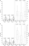
Hypoxia and angiogenesis are important factors in breast cancer progression. Little is known of hypoxia and angiogenesis in lymph node metastases of breast cancer. The aim of this study was to quantify hypoxia, by hypoxia-induced marker expression levels, and angiogenesis, by endothelial cell proliferation, comparing primary breast tumours and axillary lymph node metastases. Tissue sections of the primary tumour and a lymph node metastasis of 60 patients with breast cancer were immunohistochemically stained for the hypoxia-markers carbonic anhydrase 9 (CA9), hypoxia-inducible factor-1alpha (Hif-1alpha) and DEC-1 and for CD34/Ki-67. Endothelial cell proliferation fraction (ECP%) and tumour cell proliferation fraction (TCP%) were assessed. On haematoxylin-eosin stain, the growth pattern and the presence of a fibrotic focus were assessed. Hypoxia-marker expression, ECP% and TCP% in primary tumours and in lymph node metastases were correlated to each other and to clinico-pathological variables. Median ECP% and TCP% in primary tumours and lymph node metastases were comparable (primary tumours: ECP%=4.02, TCP%=19.54; lymph node metastases: ECP%=5.47, TCP%=21.26). ECP% correlated with TCP% (primary tumours: r=0.63, P<0.001; lymph node metastases: r=0.76, P<0.001). CA9 and Hif-1alpha expression were correlated (primary tumours P=0.005; lymph node metastases P<0.001). In primary tumours, CA9 and Hif-1alpha expression were correlated with DEC-1 expression (P=0.05), presence of a fibrotic focus (P<0.007) and mixed/expansive growth pattern (P<0.001). Primary tumours and lymph node metastases with CA9 or Hif-1alpha expression had a higher ECP% and TCP% (P<0.003); in primary tumours, mixed/expansive growth pattern and fibrotic focus were characterised by higher ECP% (P=0.03). Furthermore, between primary tumours and lymph node metastases a correlation was found for ECP%, TCP%, CA9 and Hif-1alpha expression (ECP% r=0.51, P<0.001; TCP r=0.77, P<0.001; CA9 and Hif-1alpha P<0.001). Our data demonstrate that the growth of breast cancer lymph node metastases is angiogenesis dependent and that angiogenesis and hypoxia in the primary tumour predict angiogenesis and hypoxia in the lymph node metastases. Together with previous findings in breast cancer liver metastases, which grow in 96% of cases angiogenesis independently, these data suggest that both the intrinsic growth characteristics and angiogenic potential of breast cancer cells and the site-specific tumour microenvironment determine angiogenesis and hypoxia in breast cancer.
Figures


Similar articles
-
Comparison of molecular determinants of angiogenesis and lymphangiogenesis in lymph node metastases and in primary tumours of patients with breast cancer.J Pathol. 2007 Sep;213(1):56-64. doi: 10.1002/path.2211. J Pathol. 2007. PMID: 17674348
-
Early distant relapse in "node-negative" breast cancer patients is not predicted by occult axillary lymph node metastases, but by the features of the primary tumour.J Pathol. 2001 Apr;193(4):442-9. doi: 10.1002/path.829. J Pathol. 2001. PMID: 11276002
-
Levels of hypoxia-inducible factor-1alpha independently predict prognosis in patients with lymph node negative breast carcinoma.Cancer. 2003 Mar 15;97(6):1573-81. doi: 10.1002/cncr.11246. Cancer. 2003. PMID: 12627523
-
Targeting tumour hypoxia in breast cancer.Eur J Cancer. 2008 Dec;44(18):2766-73. doi: 10.1016/j.ejca.2008.09.025. Epub 2008 Nov 5. Eur J Cancer. 2008. PMID: 18990559 Review.
-
Significance and therapeutic implications of endothelial progenitor cells in angiogenic-mediated tumour metastasis.Crit Rev Oncol Hematol. 2016 Apr;100:177-89. doi: 10.1016/j.critrevonc.2016.02.010. Epub 2016 Feb 18. Crit Rev Oncol Hematol. 2016. PMID: 26917455 Review.
Cited by
-
The Multifaceted Effects of Breast Cancer on Tumor-Draining Lymph Nodes.Am J Pathol. 2021 Aug;191(8):1353-1363. doi: 10.1016/j.ajpath.2021.05.006. Epub 2021 May 24. Am J Pathol. 2021. PMID: 34043978 Free PMC article. Review.
-
HIFs, angiogenesis, and metabolism: elusive enemies in breast cancer.J Clin Invest. 2020 Oct 1;130(10):5074-5087. doi: 10.1172/JCI137552. J Clin Invest. 2020. PMID: 32870818 Free PMC article. Review.
-
Relationships between hypoxia markers and the leptin system, estrogen receptors in human primary and metastatic breast cancer: effects of preoperative chemotherapy.BMC Cancer. 2010 Jun 22;10:320. doi: 10.1186/1471-2407-10-320. BMC Cancer. 2010. PMID: 20569445 Free PMC article.
-
Enhanced glioma-targeting and stability of LGICP peptide coupled with stabilized peptide DA7R.Acta Pharm Sin B. 2018 Jan;8(1):106-115. doi: 10.1016/j.apsb.2017.11.004. Epub 2018 Jan 3. Acta Pharm Sin B. 2018. PMID: 29872627 Free PMC article.
-
The potential of hypoxia markers as target for breast molecular imaging--a systematic review and meta-analysis of human marker expression.BMC Cancer. 2013 Nov 10;13:538. doi: 10.1186/1471-2407-13-538. BMC Cancer. 2013. PMID: 24206539 Free PMC article.
References
-
- Aranda F, Laforga J (1996) Microvessel quantitation in breast ductal invasive carcinoma. Correlation with proliferative activity, hormonal receptors and lymph node metastases. Pathol Res Pract 192: 124–129 - PubMed
-
- Arapandoni-Dadioti P, Giatromanolaki A, Trihia H, Harris AL, Koukourakis M (1999) Angiogenesis in ductal breast carcinoma. Comparison of microvessel density between primary tumour and lymph node metastasis. Cancer Lett 137: 145–150 - PubMed
-
- Blackwell K, Dewhirst M, Liotcheva V, Snyder S, Broadwater G, Bentley R, Lal A, Riggins G, Anderson S, Vredenburgh J, Proia A, Harris LN (2004) HER-2 gene amplification correlates with higher levels of angiogenesis and lower levels of hypoxia in primary breast tumors. Clin Cancer Res 10: 4083–4088 - PubMed
-
- Bos R, Van Der Groep P, Greijer A, Shvarts A, Meijer S, Pinedo H, Semenza GL, Van Diest PJ, Van Der Wall E (2003) Levels of hypoxia-inducible factor-1alpha independently predict prognosis in patients with lymph node negative breast carcinoma. Cancer 97: 1573–1581 - PubMed
Publication types
MeSH terms
LinkOut - more resources
Full Text Sources
Medical
Research Materials

