

Review
doi: 10.1038/sj.emboj.7600860.
Epub 2005 Oct 27.
The amyloid-beta precursor protein: integrating structure with biological function
Affiliations
- PMID: 16252002
- PMCID: PMC1356301
- DOI: 10.1038/sj.emboj.7600860
Item in Clipboard
Review
The amyloid-beta precursor protein: integrating structure with biological function
EMBO J.
.
Abstract
Proteolytic processing of the amyloid-beta precursor protein (APP) generates the Abeta amyloid peptide of Alzheimer's disease. The biological function of APP itself remains, however, unclear. In the current review, we study in detail the different subdomains of APP and try to assign functional significance to particular structures identified in the protein.
Figures
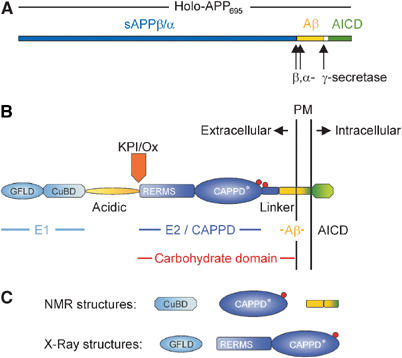
(A) Schematic representation of the holoprotein of human APP695 including the relative position of the α-, β- and γ-secretase cleavage sites. (B) Domain organization of APP: the E1 region consists of the N-terminal growth factor-like domain (GFLD) and the following copper-binding domain (CuBD). The E1 region is linked via the acidic region to the carbohydrate domain, which contains the two N-glycosylation sites of the ectodomain (red spheres). The carbohydrate domain can be subdivided into the E2 domain, also called central APP domain (CAPPD), and a linker or juxtamembrane domain. The carbohydrate domain is followed by the transmembrane and the APP intracellular domain (AICD). Aβ indicates the amyloid β-peptide sequence. The Kunitz-type protease inhibitor domain (KPI), which is present in APP751 and APP770, and the Ox2 sequence, which is present in APP770, are shown above their insertion site. (C) Known stable structures of APP.
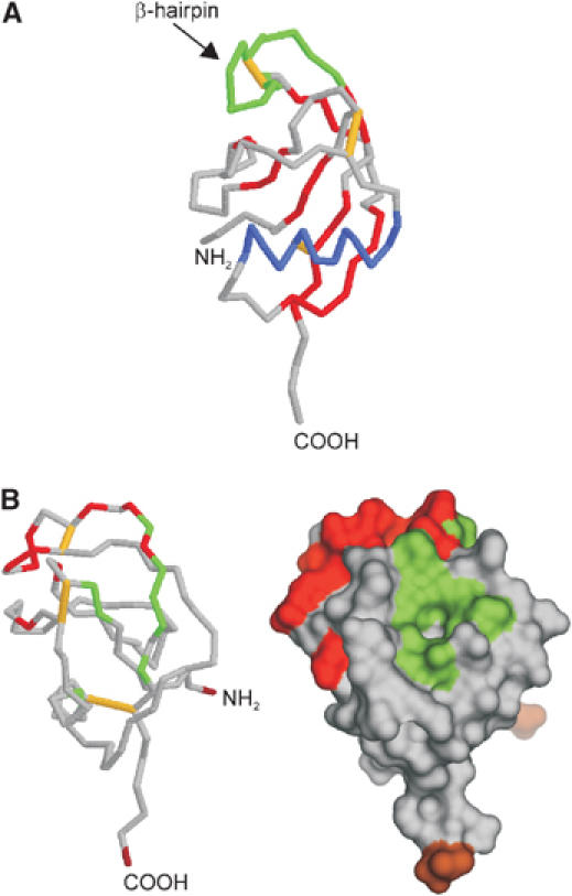
Growth factor-like domain (PDB:1MWP). (A) Backbone diagram: disulfide bridges are shown in yellow, β-sheets in red, the α-helix in blue and the β-hairpin loop in green. (B) Surface representation: the N- and C-termini are colored in brown, the hydrophobic surface patch in green and the HSPG-binging region is shown in red (note that the structure is rotated around the vertical axis by 90° compared to (A)).
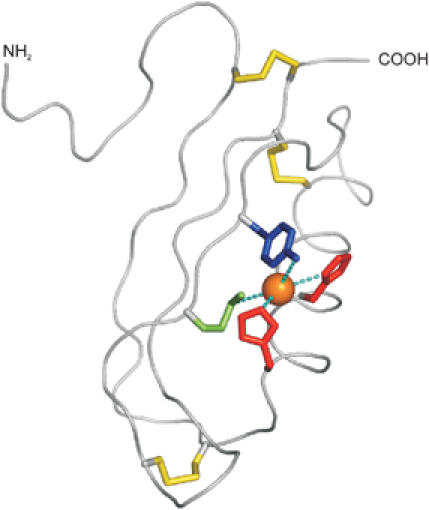
Copper-binding domain (PDB:1OWT). Backbone trace of the copper-binding domain of APP with disulphide bridges indicated in yellow. An orange sphere indicates the approximate position of a Cu(I) ion modeled to adopt a tetrahedral coordination geometry between His-147 (red), His-151 (red), Tyr-168 (blue) and Met-170 (green).
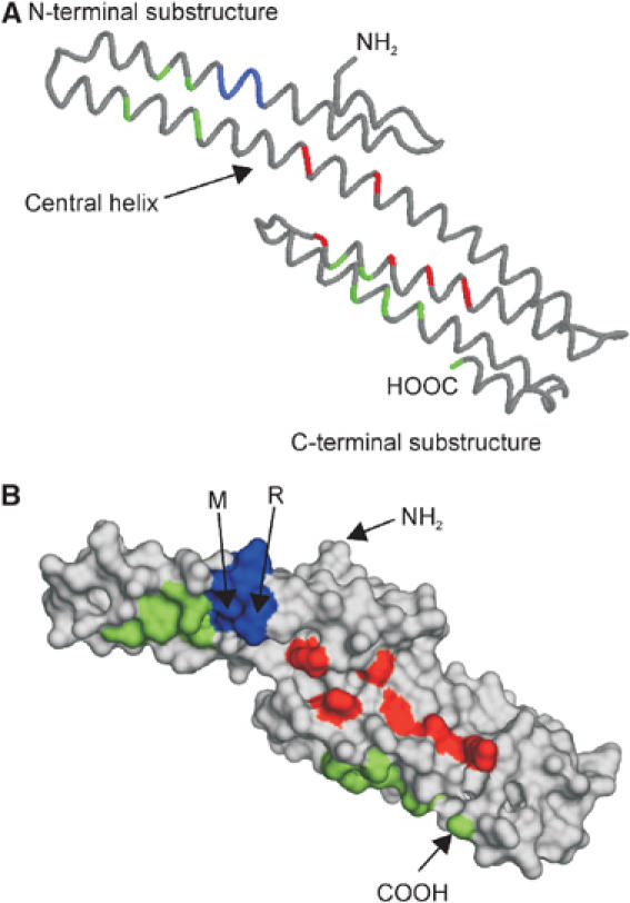
Monomeric E2 domain/central APP domain (CAPPD) (PDB:1RW6). (A) Backbone diagram: position of RERMS sequence is marked in blue, amino-acid residues forming the two hydrophobic patches are colored in green and the conserved amino-acid residues of the HSPG-binding site in red. (B) Surface representation of (A). The N- and C-termini as well as the underlined amino-acid residues of the R ERM S sequence, which participate in dimerization, are labeled.

Solution structure of Aβ42 (A) in 40% trifluorethanol (PDB:1AML) and (B) in 80% hexafluoroisopropanol (PDB:1IYT). Three models out of 20 and 10 are depicted. Note that the structure of Aβ42 is less stable in 40% trifluorethanol.
References
-
- Abramov AY, Canevari L, Duchen MR (2004) Calcium signals induced by amyloid beta peptide and their consequences in neurons and astrocytes in culture. Biochim Biophys Acta 1742: 81–87 - PubMed
-
- Ando K, Iijima KI, Elliott JI, Kirino Y, Suzuki T (2001) Phosphorylation-dependent regulation of the interaction of amyloid precursor protein with Fe65 affects the production of beta-amyloid. J Biol Chem 276: 40353–40361 - PubMed
-
- Annaert W, De Strooper B (1999) Presenilins: molecular switches between proteolysis and signal transduction. Trends Neurosci 22: 439–443 - PubMed
-
- Annaert W, De Strooper B (2002) A cell biological perspective on Alzheimer's disease. Annu Rev Cell Dev Biol 18: 25–51 - PubMed
Publication types
MeSH terms
Substances
LinkOut - more resources
Full Text Sources
Molecular Biology Databases
