. 2005 Dec 7;24(23):3985-95.
doi: 10.1038/sj.emboj.7600858. Epub 2005 Oct 27.

Structure of the split PH domain and distinct lipid-binding properties of the PH-PDZ supramodule of alpha-syntrophin

Affiliations

Structure of the split PH domain and distinct lipid-binding properties of the PH-PDZ supramodule of alpha-syntrophin

Jing Yan et al. EMBO J. .

Abstract

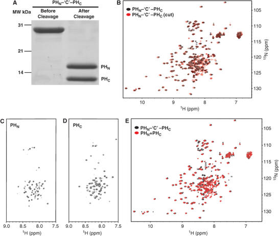
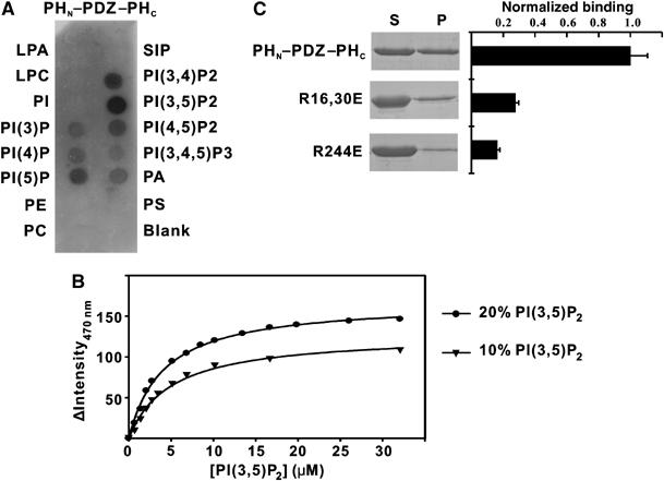
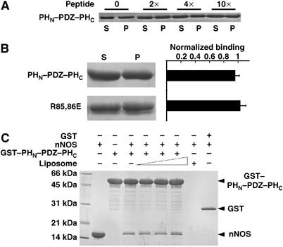
Pleckstrin homology (PH) domains play diverse roles in cytoskeletal dynamics and signal transduction. Split PH domains represent a unique subclass of PH domains that have been implicated in interactions with complementary partial PH domains 'hidden' in many proteins. Whether partial PH domains exist as independent structural units alone and whether two halves of a split PH domain can fold together to form an intact PH domain are not known. Here, we solved the structure of the PH(N)-PDZ-PH(C) tandem of alpha-syntrophin. The split PH domain of alpha-syntrophin adopts a canonical PH domain fold. The isolated partial PH domains of alpha-syntrophin, although completely unfolded, remain soluble in solution. Mixing of the two isolated domains induces de novo folding and yields a stable PH domain. Our results demonstrate that two complementary partial PH domains are capable of binding to each other to form an intact PH domain. We further showed that the PH(N)-PDZ-PH(C) tandem forms a functionally distinct supramodule, in which the split PH domain and the PDZ domain function synergistically in binding to inositol phospholipids.


Figures

Figure 1
Figure 1
Comparison of the split PH and PDZ domains in the PHN–PDZ–PHC tandem and in their respective isolated state. (A) Schematic diagram showing the domain organization of syntrophins. (B) Superposition plots of 1H, 15N HSQC spectra of the PHN–PDZ–PHC tandem (black), the isolated PDZ domain (green), and the joint PHN–PHC domain (red). (C) Plot of chemical shift changes as a function of residue number of the split PH and PDZ domains in the PHN–PDZ–PHC tandem and in their respective isolated forms. The combined 1H and 15N chemical shift changes are defined as: formula image 
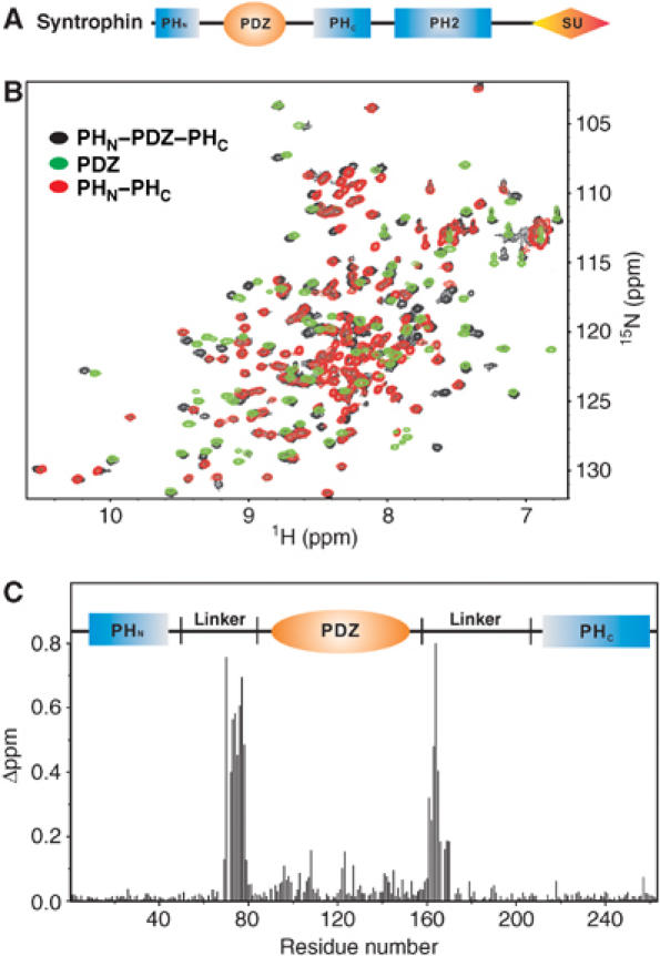
ΔδHN and ΔδN represent chemical shift differences of amide proton and nitrogen chemical shifts between the PHN–PDZ–PHC tandem and the isolated PH and PDZ domains, respectively. The scaling factor (αN) used to normalize the 1H and 15N chemical shifts is 0.17. The domain organization of the PHN–PDZ–PHC tandem is indicated at the top of the plot.
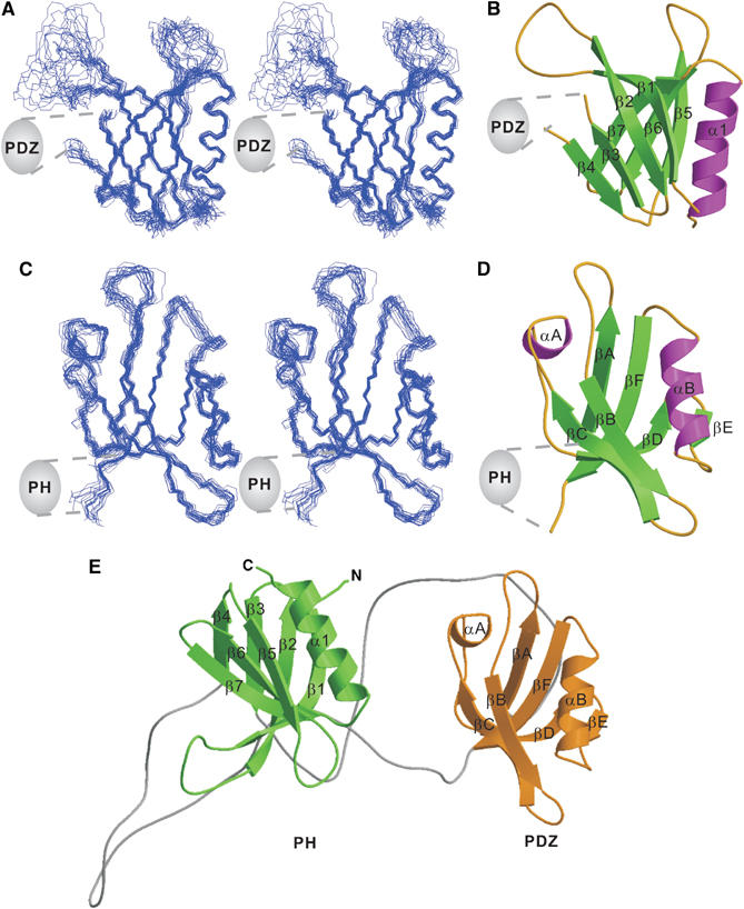
Figure 2
Figure 2
Structure of the PHN–PDZ–PHC tandem. (A) Stereo view showing the backbones of 20 superimposed NMR-derived structures of the split PH domain from the PHN–PDZ–PHC tandem. Since the orientation between the split PH domain and the PDZ domain in the PHN–PDZ–PHC tandem is not fixed, we chose to present the structures of each domain separately. (B) Ribbon diagram of a representative NMR structure of the split PH domain. The insertion of the PDZ domain in the β3/β4-loop of the split PH domain is indicated. (C) Stereo view plot of 20 superimposed NMR structures of the PDZ domain from the PHN–PDZ–PHC tandem. (D) Ribbon diagram drawing of the PDZ domain structure. The split PH domain connected to the βA and βF of the domain is indicated. (E) Ribbon diagram drawing of a representative NMR structure of the PHN–PDZ–PHC tandem. In this drawing, the split PH domain is in green, and the PDZ domain is in gold.
Figure 3
Figure 3
Folding and interaction of the two isolated, partial PH domain fragments. (A) SDS–PAGE showing the purification of the joined PHN–‘C'–PHC domain and cleavage of the domain into PHN and PHC fragments. (B) Overlay plot of the 1H, 15N HSQC spectra of the joined PHN–‘C'–PHC (black) and its protease cleaved form (red). 1H, 15N HSQC spectra of the isolated PHN (C) and PHC (D) fragments. (E) Overlay plot of the 1H, 15N HSQC spectra of the joined PHN–‘C'–PHC (black) and the 1:1 mixture of the two halves of the split PH domain (red).
Figure 4
Figure 4
The PHN–PDZ–PHC supramodule binds to brain liposomes with enhanced avidity. (A) Schematic diagrams showing the various forms of proteins purified for liposome-binding assays. (B) Brain liposome-binding assays of the PHN–PDZ–PHC tandem and its fragments. ‘S' and ‘P' denote proteins recovered in the supernatants and pellets, respectively, in the centrifugation-based liposome-binding assays. (C) Dose-dependent binding between the PHN–PDZ–PHC tandem and the brain liposome. In this assay, the amount of the PHN–PDZ–PHC tandem is fixed at 10 μM, and the concentration of liposome varies. The liposome-bound PHN–PDZ–PHC tandem is recovered in the pellet in the binding assay. (D) Lipid binding of the two mutants of the PHN–PDZ–PHC tandem. In these two mutants, the PDZ domain was placed either at the front or after the split PH domain. In these mutants, the PH and the PDZ domains were connected by a flexible ‘Gly–Ser–Gly–Gly–Ser–Gly–Gly–Ser–Gly–Ser' linker. (E) Lipid binding of a PHN–PDZ–PHC tandem mutant with the two connecting linkers shortened to six residues. Amino-terminal His6-tagged proteins were used in all of the brain liposome-binding assays.
Figure 5
Figure 5
Lipid-binding specificity of the PHN–PDZ–PHC supramodule using phospholipid strip overlay assay. (A) Binding of the PHN–PDZ–PHC tandem to the phospholipids spotted on a cellulose membrane. The amount of lipid per spot was 100 pmol. Abbreviations for the lipids: S1P: sphingosine-1-phosphate; LPA: lysophosphatidic acid; LPC: lysophosphocholine; PE: phosphatidylethanolamine; PS: phosphatidylserine; PA: phosphatidic acid; PC: phosphatidylcholine; PtdIns: phosphatidylinositol. The amount of protein used in the assay is 0.5 μg/ml. (B) Binding isotherms of PHN–PDZ–PHC with PC/PS liposomes containing either 10 or 20% PI(3,5)P2. (C) Identification of the residues in the split PH domain of the PHN–PDZ–PHC tandem that are involved in the lipid binding.
Figure 6
Figure 6
The PDZ ligands do not affect lipid binding of the PHN–PDZ–PHC tandem. (A) Addition of excess amounts (up to ∼10 equivalents) of a PDZ-binding peptide to the liposome-binding assay buffer did not alter the lipid-binding capacity of the PHN–PDZ–PHC tandem. The amino-acid sequence of the PDZ ligand peptide is: KLSSIESDV. (B) Mutation of the two positively charged residues (Arg85 and Arg86) in the carboxyl group-binding loop of the target recognition groove of the PDZ domain did not change lipid-binding capacity of the PHN–PDZ–PHC tandem. (C) Binding of lipids to the PHN–PDZ–PHC tandem does not change the binding of PDZ domain to its ligand, the nNOS PDZ domain. Three different liposome concentrations (0.4, 0.6, and 0.8 mg/ml) were tested in this assay.

Similar articles

Cited by

References

    1. Adams ME, Butler MH, Dwyer TM, Peters MF, Murnane AA, Froehner SC (1993) Two forms of mouse syntrophin, a 58 kd dystrophin-associated protein, differ in primary structure and tissue distribution. Neuron 11: 531–540 - PubMed
    1. Adams ME, Kramarcy N, Krall SP, Rossi SG, Rotundo RL, Sealock R, Froehner SC (2000) Absence of alpha-syntrophin leads to structurally aberrant neuromuscular synapses deficient in utrophin. J Cell Biol 150: 1385–1398 - PMC - PubMed
    1. Adams ME, Mueller HA, Froehner SC (2001) In vivo requirement of the alpha-syntrophin PDZ domain for the sarcolemmal localization of nNOS and aquaporin-4. J Cell Biol 155: 113–122 - PMC - PubMed
    1. Ahn AH, Kunkel LM (1995) Syntrophin binds to an alternatively spliced exon of dystrophin. J Cell Biol 128: 363–371 - PMC - PubMed
    1. Ahn AH, Yoshida M, Anderson MS, Feener CA, Selig S, Hagiwara Y, Ozawa E, Kunkel LM (1994) Cloning of human basic A1, a distinct 59-kDa dystrophin-associated protein encoded on chromosome 8q23–24. Proc Natl Acad Sci USA 91: 4446–4450 - PMC - PubMed

Publication types