. 2005 Nov 16;24(22):3869-80.
doi: 10.1038/sj.emboj.7600856. Epub 2005 Oct 27.

Critical role of novel Thr-219 autophosphorylation for the cellular function of PKCtheta in T lymphocytes

Affiliations

Critical role of novel Thr-219 autophosphorylation for the cellular function of PKCtheta in T lymphocytes

Nikolaus Thuille et al. EMBO J. .

Abstract

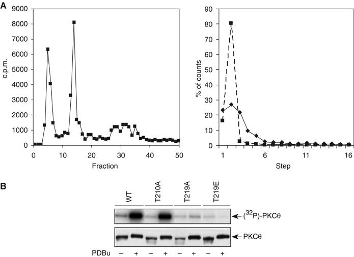
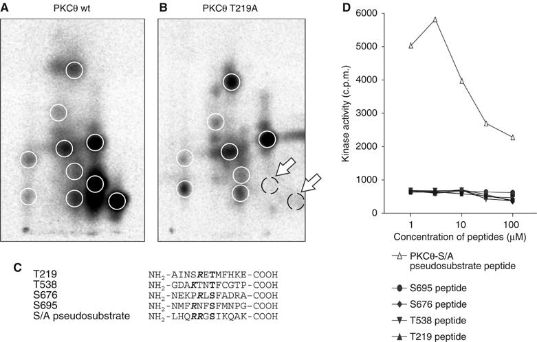
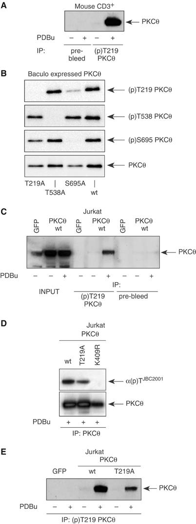
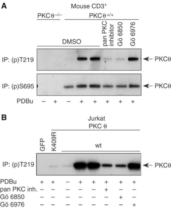
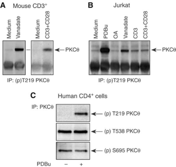
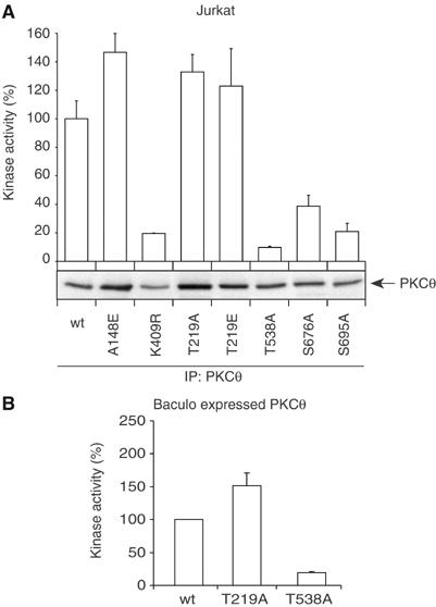
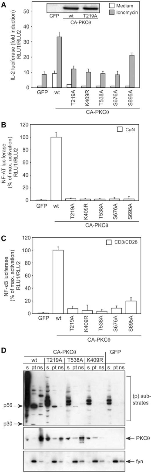
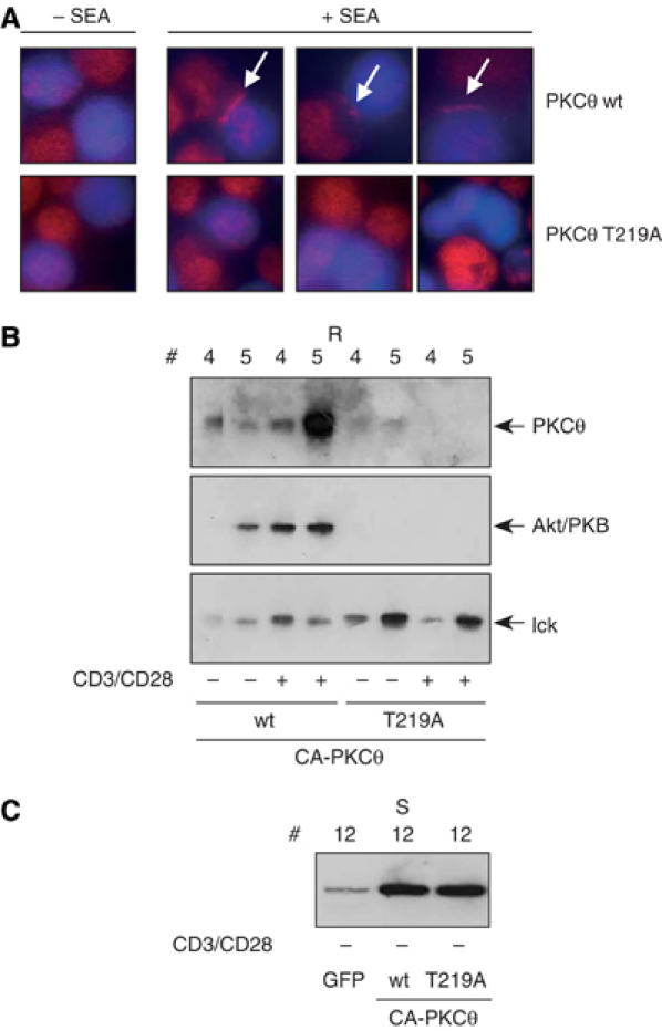
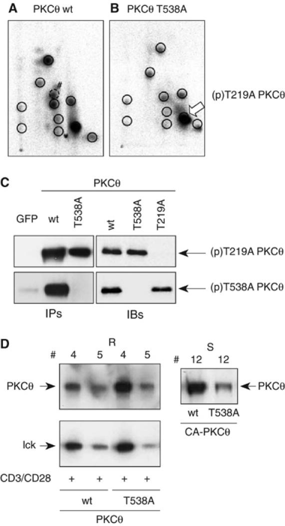
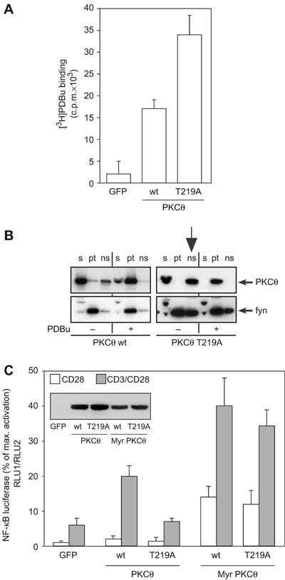
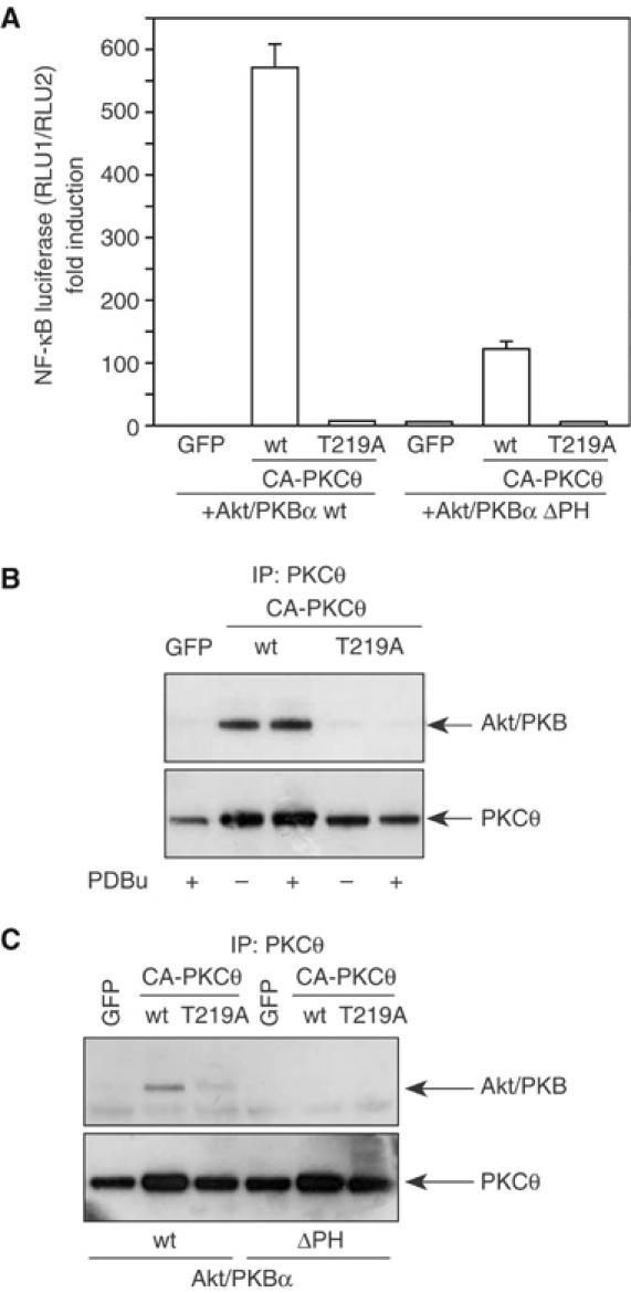
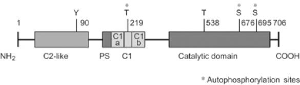
Phosphopeptide mapping identified a major autophosphorylation site, phospho (p)Thr-219, between the tandem C1 domains of the regulatory fragment in protein kinase C (PKC)theta. Confirmation of this identification was derived using (p)Thr-219 antisera that reacted with endogenous PKCtheta in primary CD3+ T cells after stimulation with phorbol ester, anti-CD3 or vanadate. The T219A mutation abrogated the capacity of PKCtheta to mediate NF-kappaB, NF-AT and interleukin-2 promoter transactivation, and reduced PKCtheta's ability in Jurkat T cells to phosphorylate endogenous cellular substrates. In particular, the T219A mutation impaired crosstalk of PKCtheta with Akt/PKBalpha in NF-kappaB activation. Yet, this novel (p)Thr-219 site did not affect catalytic activity or second-messenger lipid-binding activity in vitro. Instead, the T219A mutation prevented proper recruitment of PKCtheta in activated T cells. The PKCthetaT219A mutant defects were largely rescued by addition of a myristoylation signal to force its proper membrane localization. We conclude that autophosphorylation of PKCtheta at Thr-219 plays an important role in the correct targeting and cellular function of PKCtheta upon antigen receptor ligation.


Figures

Figure 1
Figure 1
Dominant autophosphorylation of PKCθ. (A) PKCθ was autophosphorylated in vitro, digested with trypsin, fractions separated by HPLC and Cerenkov emissions measured (left panel). The major peaks of the digest (fractions 5, solid line and fraction 14, dashed line) were subjected to sequential release of residues by Edman degradation and released fractions assessed for β emissions measured in the presence of scintillant (right panel). (B) Transfected PKCθ wild-type or mutant cDNA constructs were immunoprecipitated with anti-tag antibodies and in vitro kinase assay performed in the presence of γ32P-ATP with or without 100 nM PDBu stimulation, as indicated. Product was analyzed by SDS–PAGE, followed by autoradiography. Note that PKCθT219A and T219E mutants were mostly unable to autophosphorylate. Equal concentration of kinase in each lane was confirmed by immunoblotting.
Figure 2
Figure 2
Thr-219 is a major contributor to autophosphorylation. Tryptic phosphopeptide maps of baculo-expressed and purified PKCθ wild-type (A) or PKCθ T219A mutant proteins (B). The circles depict the major peptides obtained from PKCθ. Note that two major phosphopeptides were missing in the mutant, as indicated by the arrows. (C) Amino-acid sequences of the peptide containing Thr-219, Thr-538, Ser-676 and Ser-695 of human PKCθ; phosphorylation site and NH2-terminal basic residues are shown in bold. (D) Phosphorylation of these substrate peptides, employing recombinant baculo-expressed PKCθ, and scintillation proximity kinase assays are shown.
Figure 3
Figure 3
Thr-219 phosphorylation in PKCθ is inducible. (A) PKCθ was immunoprecipitated from resting (−) or 20 min and 100 nM PDBu-stimulated (+) primary CD3+ mouse cells and immunoblotted with PKCθ-specific mAb. (B) Baculo-expressed and isolated PKCθ mutant proteins were tested with phosphopeptide-specific antibodies, (p)T219, (p)T538 and (p)S695 on total wild-type and mutant PKCθ proteins to confirm their phosphoepitope specificity. (C) The stoichiometry of the Thr-219 site-specific phosphorylation of PKCθ was estimated with immunoprecipitation employing the (p)Thr-219-specific antibody versus total recombinant PKCθ protein (INPUT) from PKCθ wild-type cDNA transfected cell lysates. This procedure was described in Miinea and Lienhard (2003). (D, E) Jurkat T cells, transiently transfected with GFP inert protein control, PKCθ wild type or PKCθT219A or PKCθK406R, as indicated were immunoblotted with PKCθ-specific mAb. One representative result out of three independent experiments was shown.
Figure 4
Figure 4
Thr-219 phosphorylation depends on PKCθ catalytic activity. PKCθ was immunoprecipitated from resting (−) or 20 min and 100 nM PDBu-stimulated (+) mouse CD3+ cells, purified from lymph nodes and spleen (A), and Jurkat cells (B), transfected with GFP control, PKCθ wild type or PKCθK406R as indicated. The Thr-219 phosphostatus was determined as in Figure 3, except that the cells were pretreated for 1 h with distinct PKC inhibitors (at 500 nM concentrations) or DMSO buffer control, as indicated. In contrast, the classical hydrophobic motif autophosphorylation site of PKCθ, (p)S695, was constitutively phosphorylated and induced much less upon stimulation (A, lower panel). One representative result out of three independent experiments was shown.
Figure 5
Figure 5
Phosphostatus analysis of PKCθ. Thr-219 phosphorylation of endogenous PKCθ in primary CD3+ cells (A), in Jurkat cells, transfected with PKCθ wild-type cDNA (B) and in primary human CD4+ cells (C), treated with medium alone, solid-phase CD3 and/or CD28 antibodies, 100 nM PDBu, 200 μM pervanadate or 1 μM okadaic acid for 20 min at 37°C. Similar results were obtained in at least three independent experiments.
Figure 6
Figure 6
Thr-219 is not required for the catalytic activity of PKCθ. (A) In vitro kinase assay of PKCθ immunoprecipitates from Jurkat cells transfected with PKCθ cDNA expression plasmids as indicated. The upper panel shows the ability of PKCθ to phosphorylate the pseudosubstrate peptide, and the lower panel represents the corresponding anti-PKCθ immunoblots. The kinase activity thereby has been normalized for each PKCθ mutant protein by the expression levels, and results expressed relative to that measured with the wild-type protein. (B) Similar experiment as in panel A, except that baculo-expressed and purified PKCθ protein was used as the source (P<0.005 in (A) and (B); t-test).
Figure 7
Figure 7
Thr-219 is required for the activity of PKCθ in Jurkat T cells. (A–C) Jurkat cells transfected with GFP inert protein control or CA mutants of PKCθ and CaN as indicated. Expression of the transgenes has been confirmed by immunoblotting of equal amounts of total cell lysates (inset). Luciferase activity in lysates of cells cotransfected with the indicated PKCθ expression constructs, luciferase reporter plasmid, and either left unstimulated or stimulated with 500 nM ionomycin or solid-phase CD3/CD28 antibodies for 16 h at 37°C. The measured firefly luciferase activity was normalized for transfection efficiency using the renilla luciferase activity, and results were expressed relative to that induced by the unstimulated GFP control. Values represent the average of at least three experiments carried out in duplicates (P<0.005 in (A–C); t-test). (D) Transiently transfected Jurkat T cells were subjected to biochemical fractionation and subsequent immunodetection with a broadly reactive anti-phosphoserine PKC substrate antibody (able to detect distinct phospho-Ser containing endogenous proteins): biochemical fractions corresponding to cytosol (s), membrane (pt) and cytoskeletal fractions (ns) were prepared by standard fractionation, resolved by SDS–PAGE, and the phospho-Ser PKC substrate staining of cellular proteins. Additionally, recombinant expression and relative subcellular distribution of the PKCθ mutants (in comparison to the GFP vector control) was determined. As positive evaluation for cell fractionation, immunostaining of p59fyn predominantly recognized the pt fraction. The results are representative of three experiments.
Figure 8
Figure 8
Thr-219 is required for submembranous location of PKCθ in Jurkat T cells. (A) Jurkat cells were transfected with PKCθ wild-type or T219A mutant, as indicated, and 21 h later incubated with Raji B cells (stained in blue) in the absence or presence of SEA for 15 min. Recombinant PKCθ was then stained in red by immunofluorescence. Representative images out of three independent experiments are shown. The arrows mark PKCθ enrichment in the T- and B-cell contact zone. (B, C) Jurkat T cells transfected with CA-PKCθ wild-type or CA-PKCθT219A cDNA, respectively, as indicated. Lipid raft fractions (R) and detergent-soluble fraction (S) were immunostained for PKCθ and Akt/PKB in order to differentially contrast protein lipid raft distributions of the same cells. As a marker for the raft fraction, p56lck was used for immunoblots of the same samples. A representative experiment of three independent experiments was shown.
Figure 9
Figure 9
‘Kinase-low' Thr-538 activation loop mutant is phosphorylated at Thr-219 and demonstrates intact submembranous location of PKCθ in Jurkat T cells. Tryptic phosphopeptide maps of baculo-expressed and purified PKCθ wild-type (A) or PKCθT538A mutant proteins (B). The circles depict the peptides obtained from PKCθ. The major (p)Thr-219 representing phosphopeptide was indicated by the arrow. (C) Recombinant wild-type and mutant PKCθ was immunoprecipitated (IPs—left panel) from PDBu-stimulated Jukat cells or purified from baculovirus-infected Sf21 cells (IBs—right panel) and immunoblotted with the phosphopeptide-specific antibodies, (p)T219 and (p)T538, as indicated. (D) Jurkat T cells transfected with CA-PKCθ wild-type or CA-PKCθT538A cDNA, respectively, as indicated. Lipids raft fractions (R) and detergent-soluble fraction (S) were immunostained for PKCθ and p56lck.
Figure 10
Figure 10
Thr-219 is not required for PDBu binding in Jurkat T cells. Jurkat T cells were transfected with PKCθ wild-type or PKCθT219A cDNA expression vectors, as indicated. After 21 h, cells were (A) determined for specific 3H-PDBu binding as described (Chida and Kuroki, 1983) and (B) stimulated with 100 nM PDBu for 20 min or left unstimulated, as indicated. Subcellular distribution of PKCθ (upper panel) and p59fyn (lower panel) was then determined by immunoblotting. The cell fractions were defined as the soluble (s) fraction, the particulate (pt) fraction and the Triton X-100 nonsoluble (ns) fraction. Note the constitutive enrichment of the T219A mutant in the ns fraction of nonstimulated cells (indicated by the arrow). (C) MyrPKCθT219A synergizes with CD3/CD28 in mediated NF-κB reporter activation. cDNA expression vectors encoding GFP control, wild-type and Myr-NH2-terminal fusion mutants of PKCθ, respectively, together with the NF-κB reporter plasmid have been cotransfected in Jurkat T cells. After 21 h, cells were stimulated and assayed for reporter gene expression. Expression of the PKCθ transgenes has been confirmed by immunoblotting (inset).
Figure 11
Figure 11
Thr-219 is required for PKCθ crosstalk with Akt/PKBα in T cells. (A) Jurkat T cells were transfected with CA-PKCθ and Akt/PKBα constructs, as indicated, stimulated for 16 h with 500 nM ionomycin and analyzed as in Figure 7. Statistical analysis of three independent experiments was evaluated as the means of fold induction±s.e.; P<0.005. Expression of the transgenes has been confirmed by immunoblotting (not shown). (B) Coimmunoprecipitation analysis of recombinant PKCθ and endogenous Akt/PKB. PKCθ immunoprecipitations of Jurkat T-cell lysates, transfected 21 h earlier with GFP control, CA-PKCθ or CA-PKCθT219A expression vectors (as indicated). Jurkat T cells were stimulated for 20 min at 37°C with 100 nM PDBu or left unstimulated. Afterwards, cell extracts were immunoprecipitated (IP) with an anti-PKCθ antibody. (C) Coimmunoprecipitation analysis of recombinant PKCθ and Akt/PKBα in nonstimulated Jurkat T-cell lysates, transfected 21 h earlier with GFP control, CA-PKCθ or CA-PKCθT219A and Akt/PKBα wild-type and PH domain deletion mutant (ΔPH) expression vectors (as indicated). Immunoblots were stained for PKCθ (lower panel) and Akt/PKB (upper panel), respectively. Comparable results were obtained in two independent experiments.
Figure 12
Figure 12
Cartoon of PKCθ domain structure and phosphorylation sites. The domain structure of PKCθ was schematically shown and consists of a conserved serine/threonine kinase (COOH-terminus) and a regulatory (NH2-terminus) domain. Thr-219 resides within the cystein-rich (C1) sequences of PKCθ, responsible for second-messenger lipid interaction. The established phosphorylation sites of serine (S), threonine (T), and tyrosine (Y) residues are indicated.

Similar articles

Cited by

References

    1. Baier G (2003) The PKC gene module: molecular biosystematics to resolve its T cell functions. Immunol Rev 192: 64–79 - PubMed
    1. Baier-Bitterlich G, Uberall F, Bauer B, Fresser F, Wachter H, Grunicke H, Utermann G, Altman A, Baier G (1996) PKCθ isoenzyme selective stimulation of the transcription factor complex AP-1 in T lymphocytes. Mol Cell Biol 16: 1842–1850 - PMC - PubMed
    1. Bauer B, Baier G (2002) PKC and AKT/PKB in CD4+ T-lymphocytes: new partners in TCR/CD28 signal integration. Mol Immunol 38: 1087–1099 - PubMed
    1. Bauer B, Jenny M, Fresser F, Uberall F, Baier G (2003) AKT1/PKBα is recruited to lipid rafts and activated downstream of PKC isotypes in CD3-induced T cell signaling. FEBS Lett 541: 155–162 - PubMed
    1. Bauer B, Krumbock N, Fresser F, Hochholdinger F, Spitaler M, Simm A, Uberall F, Schraven B, Baier G (2001) Complex formation and cooperation of PKCθ and Akt1/PKBα in the NF-κB transactivation cascade in Jurkat T cells. J Biol Chem 15: 15 - PubMed

Publication types