. 2005 Nov 16;24(22):3881-94.
doi: 10.1038/sj.emboj.7600853. Epub 2005 Oct 27.

Ligand-induced transrepression by VDR through association of WSTF with acetylated histones

Affiliations

Ligand-induced transrepression by VDR through association of WSTF with acetylated histones

Ryoji Fujiki et al. EMBO J. .

Retraction in

Abstract

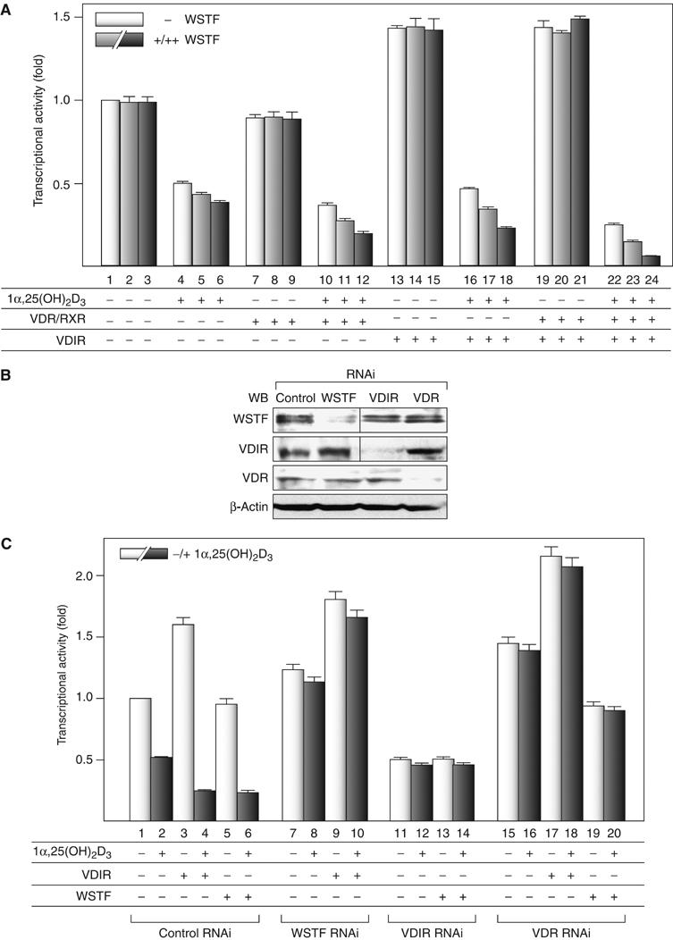
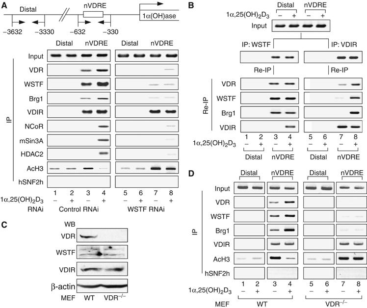
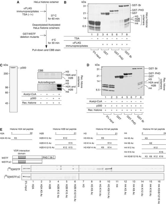
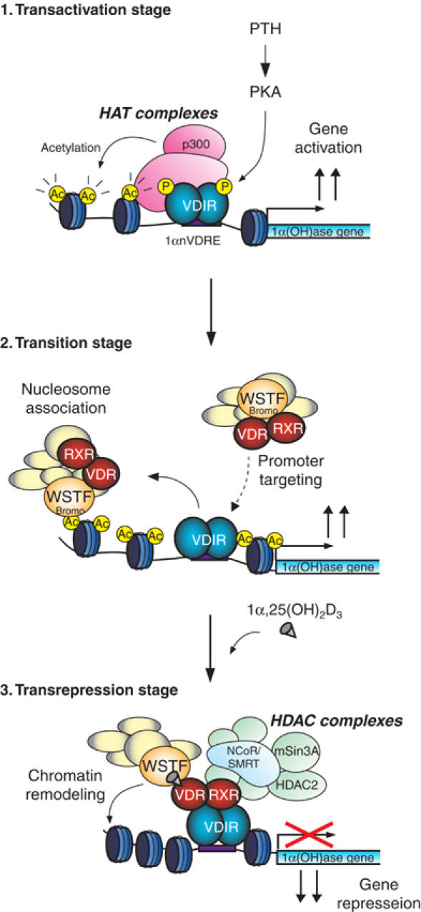
We have previously shown that the novel ATP-dependent chromatin-remodeling complex WINAC is required for the ligand-bound vitamin D receptor (VDR)-mediated transrepression of the 25(OH)D3 1alpha-hydroxylase (1alpha(OH)ase) gene. However, the molecular basis for VDR promoter association, which does not involve its binding to specific DNA sequences, remains unclear. To address this issue, we investigated the function of WSTF in terms of the association between WINAC and chromatin for ligand-induced transrepression by VDR. Results of in vitro experiments using chromatin templates showed that the association of unliganded VDR with the promoter required physical interactions between WSTF and both VDR and acetylated histones prior to VDR association with chromatin. The acetylated histone-interacting region of WSTF was mapped to the bromodomain, and a WSTF mutant lacking the bromodomain served as a dominant-negative mutant in terms of ligand-induced transrepression of the 1alpha(OH)ase gene. Thus, our findings indicate that WINAC associates with chromatin through a physical interaction between the WSTF bromodomain and acetylated his tones, which appears to be indispensable for VDR/promoter association for ligand-induced transrepression of 1alpha(OH)ase gene expression.


Figures

Figure 1
Figure 1
WSTF enhances 1α,25(OH)2D3-induced transrepression of 1α(OH)ase gene expression, but not transactivation by VDIR. (A) Coordinate transrepression of the 1α(OH)ase gene by VDR, WSTF and VDIR in a luciferase reporter assay. MCF7 cells were transfected with a luciferase reporter gene expression vector containing 1αnVDRE (× 2) driven by a TATA promoter (0.4 μg), pML-CMV (2 ng), and either pSG5-rat VDR and pSG5-rat RXRα (0.2 μg each), pcDNA3-VDIR (0.1 μg), pcDNA3-WSTF (0.1[+], 0.3[++] μg), or combinations thereof in the presence or absence of 1α,25(OH)2D3 (10−8 M) (Kitagawa et al, 2003; Murayama et al, 2004). Bars in each graph show the fold change in luciferase activity relative to basal activity obtained in the absence of ligand. All values are means±s.d. for at least three independent experiments. (B) Gene-specific knockdown of WSTF, VDIR or VDR by RNAi was confirmed by Western blots using anti-WSTF, -VDIR, -VDR and β-actin (as a control). Whole-cell extracts were prepared from MCF7 cells transfected with 0.3 μg of double-stranded siRNA and further cultured for 48 h. (C) Effect of gene-specific knockdown of endogenous factors, WSTF, VDIR and VDR on 1α(OH)ase gene expression in a luciferase reporter assay. MCF7 cells were transfected with 0.3 μg of the indicated siRNAs, 48 h after the transfection luciferase reporter gene containing 1α(OH)ase native promoter was transfected again into the cells. Luciferase activity was assessed after 12 h culture in the presence or absence of 1α,25(OH)2D3 (10−8 M).
Figure 2
Figure 2
WSTF interacts with VDIR through 1α,25(OH)2D3-bound VDR. (A) Exogenous WSTF interacted with exogenous VDIR and endogenous corepressors in an 1α,25(OH)2D3-dependent manner in vivo. MCF7 cells were transfected with 0.3 μg of WSTF, VDR and VDIR expression vector. The panels show results of immunoprecipitation with anti-VDR, -VDIR or -FLAG (WSTF) antibodies, followed by Western blot analysis using the indicated antibodies. (B) WSTF associates with HDAC activity in a ligand-dependent manner. MCF7 cells were transfected with pcDNA3 or FLAG-WSTF/pcDNA3 and the extracted cell lysates were then immunoprecipitated with anti-FLAG M2 resin. HDAC activity in the immunoprecipitates was measured by fluorometric detection using an HDAC assay kit. (C) 1α,25(OH)2D3-dependent interaction between endogenous WSTF and VDIR in vivo. MCF7 cells cultured with or without 1α,25(OH)2D3 for 12 h were subjected to immunoprecipitation with anti-WSTF or anti-VDIR antibodies. Immunoprecipitates were Western blotted with specific antibodies as shown on the left. (D) SDS–PAGE gels of a series of GST-fused WSTF deletion mutants (left panel) and recombinant VDR (right panel) were visualized by CBB staining. Recombinant proteins were expressed in Escherichia coli and purified by affinity chromatography. (E) GST pull-down assay. Schematic diagrams of the WSTF deletion mutants used are illustrated. 35S-labeled VDR translated in vitro was incubated with deletion mutants immobilized onto glutathione-Sepharose beads in the presence or absence of 1α,25(OH)2D3 (10−6 M). Bound proteins were resolved by SDS–PAGE followed by autoradiography (upper panel). Autoradiographs show 35S-labeled VDIR, preincubated with (lower panel) or without (middle panel) cold recombinant VDR, bound to the GST-fused mutants immobilized on beads (Murayama et al, 2004).
Figure 3
Figure 3
VDR is indispensable for ligand-induced promoter assembly of the WINAC and HDAC corepressor complex. (A) Recruitment of VDR, WSTF, VDIR and other coregulators to the 1α(OH)ase gene promoter in vivo, as shown by ChIP analysis. Soluble chromatin was prepared from MCF7 cells treated with 1α,25(OH)2D3 (10−8 M) for 45 min and immunoprecipitated with the indicated antibodies. Extracted DNA samples were amplified using primer pairs that covered the 1α(OH)ase negative VDRE region (1αnVDRE) (Kitagawa et al, 2003; Murayama et al, 2004) or a distal region (3 kb upstream of 1αnVDRE) as a control. (B) Recruitment of ligand-free VDR/WSTF complexes to the 1α(OH)ase gene promoter, shown by the re-chromatin immunoprecipitation (Re-ChIP) assay. Chromatin prepared from MCF7 cells cultured in the presence or absence of 1α,25(OH)2D3 (10−8 M) for 45 min was subjected to the ChIP procedure with the indicated antibodies and immunoprecipitated using the antibodies as shown on the left. (C) Cessation of VDR expression in VDR−/− MEF cells was confirmed by Western blot analysis. VDR−/− and wild-type (WT) MEF cells were generated from VDR−/− knockout mouse embryos and WT littermates (E 13.5). (D) Effect of VDR disruption in recruitment of WSTF and VDIR to the 1α(OH)ase gene promoter in vivo by ChIP analysis. Soluble chromatin was prepared from VDR−/− and WT MEF cells treated with 1α,25(OH)2D3 (10−8 M) for 45 min and subjected to the ChIP procedure as described in panel A.
Figure 4
Figure 4
Chromatin structures are required to target unliganded VDR to the 1α(OH)ase promoter. (A) SDS–PAGE analysis of purified HeLa histone octamers, recombinant histone octamers, recombinant Drosophila NAP1 (dNAP1) and Drosophila ACF (dACF) complexes. HeLa histone octamers were purified from HeLa nuclear pellets by traditional hydroxylapatite chromatography, as described in Materials and methods. Each component of the recombinant histone octamer, H2A, H2B, H3 and H4, was expressed in an insoluble form in E. coli and extracted with guanidine hydrochloride. Extracted crude proteins were further purified by traditional gel filtration and ion exchange chromatography, as described previously (Luger et al, 1999). Affinity-tagged recombinant dNAP1 and dACF complex components (FLAG-dAcf1 and dISWI) were expressed in Sf9 cells by infection with recombinant baculoviruses and purified by affinity chromatography as described in Materials and methods. (B) Chromatin template containing the 1α(OH)ase gene promoter immobilized to streptavidin beads. Schematic representation of the DNA template containing the 1α(OH)ase gene promoter is illustrated above. Chromatinized template reconstituted in vitro was confirmed using the standard MNase digestion assay. The 123 bp ladder DNA was used as a size marker. (C) Immobilized template was subjected to Western blot analysis with an anti-FLAG antibody. To eliminate possible contamination by recombinant dACF complexes, immunoblotting of the beads using anti-FLAG, acetylated histone H3 and unmodified histone H3 (as a control) antibodies was performed after extensive washing with high-salt buffer. (D) Schematic diagram of the in vitro-immobilized DNA/chromatin template assay. (E) Stabilization of the ligand-free VDR/WSTF complex on the 1α(OH)ase promoter required chromatin structure in vitro. Whole-cell extracts from MCF7 cells stably expressing FLAG-WSTF treated with or without 1α,25(OH)2D3 (10−8 M) were mixed with immobilized templates. The template beads were then concentrated using a magnet and analyzed by Western blotting using the indicated antibodies.
Figure 5
Figure 5
WSTF bromodomains in its C-terminal region interact with acetylated histone octamers. (A) Schematic diagram of a histone binding assay. HDAC immunoprecipitate was prepared using an anti-FLAG antibody from whole-cell extracts of 1α,25(OH)2D3-treated MCF7 cells transiently expressing FLAG-WSTF (see Figure 2B). (B) An in vitro histone binding assay showed that the interaction between WSTF bromodomains and HeLa histone octamers is decreased after deacetylation of the HeLa histone octamers. HeLa histone octamers were incubated with HDAC complexes, mixed with GST-fused WSTF bromodomain and PHD finger regions and then immobilized onto glutathione-Sepharose beads. Bound materials were eluted from the resin and resolved by 18% SDS–PAGE. Proteins were visualized by CBB staining. (C) SDS–PAGE analysis of recombinant p300 and in vitro histone acetylation of recombinant histone octamers. Affinity-tagged recombinant p300 was expressed in Sf9 cells using a baculovirus system and purified by affinity chromatography as described in Materials and methods. Histone octamers were acetylated in vitro by p300 with radiolabeled acetyl-CoA and the gels were visualized by CBB staining following exposure to film. (D) Interaction between recombinant histone octamers and WSTF bromodomains is enhanced after in vitro histone acetylation by p300. Recombinant histone octamers were preincubated with p300 in the absence (upper panel) or presence (lower panel) of acetyl-CoA and subjected to histone binding assay. (E) Site-specific recognition between the WSTF bromodomain and histones with tail modification. Schematic diagrams of the WSTF deletion mutants used are illustrated (upper panel). 35S-labeled WSTF and a WSTFΔC mutant translated in vitro were incubated with a series of acetylated N-terminal histone tails immobilized onto streptavidin beads. Histone tail peptides were tested for WSTF binding (middle panel). Bound WSTF was resolved by SDS–PAGE, followed by autoradiography (lower panel).
Figure 6
Figure 6
The WSTF C-terminal region is indispensable for the promoter targeting of ligand-unbound VDR and for VDR-mediated transrepression of the 1α(OH)ase gene. (A) WSTFΔC shows no binding to acetylated histone H3. MCF7 cells transfected with FLAG-tagged WSTF, FLAG-tagged WSTFΔC or pcDNA3 vector as a control were lysed and subjected to immunoprecipitation with anti-FLAG. Immunoprecipitates were Western blotted with indicated antibodies (lower panel). (B) Histone acetylation-dependent recruitment of WSTF to the 1αnVDRE region. MCF7 cells transfected with FLAG-tagged WSTF or FLAG-tagged WSTFΔC were treated with either 1α,25(OH)2D3 (10−8 M) for 45 min or TSA (10−7 M) for 120 min and then subjected to ChIP analysis. (C) A WSTF mutant with a deleted C-terminal bromodomain and PHD finger (WSTFΔC) exerted a partial dominant-negative effect on the ligand-induced transrepression function of VDR. The amounts of each transfected plasmid are described in Figure 1A.
Figure 7
Figure 7
Model demonstrating the role of WINAC in the ligand-induced transrepression function of VDR at the 1α(OH)ase gene promoter. p300 is recruited to VDIR, which was phosphorylated via PKA signaling, and it acetylates the nucleosomes around the 1α(OH)ase gene promoter region (transactivation stage). WINAC, along with VDR, sequentially targets VDIR through interaction between unliganded VDR and VDIR, and is retained on the acetylated promoter via the WSTF bromodomain. VDR becomes receptive to 1α,25(OH)2D3 binding (transition stage). Upon 1α,25(OH)2D3 binding, HDAC corepressor complexes are recruited to the ligand-bound VDR/VDIR complex, and they then deacetylate the nucleosomes. WINAC then exerts its ATP-dependent chromatin-remodeling activity (transrepression stage).

Similar articles

Cited by

References

    1. Agalioti T, Lomvardas S, Parekh B, Yie J, Maniatis T, Thanos D (2000) Ordered recruitment of chromatin modifying and general transcription factors to the IFN-beta promoter. Cell 103: 667–678 - PubMed
    1. Brenza HL, Kimmel-Jehan C, Jehan F, Shinki T, Wakino S, Anazawa H, Suda T, DeLuca HF (1998) Parathyroid hormone activation of the 25-hydroxyvitamin D3-1alpha-hydroxylase gene promoter. Proc Natl Acad Sci USA 95: 1387–1391 - PMC - PubMed
    1. Cote J, Peterson CL, Workman JL (1998) Perturbation of nucleosome core structure by the SWI/SNF complex persists after its detachment, enhancing subsequent transcription factor binding. Proc Natl Acad Sci USA 95: 4947–4952 - PMC - PubMed
    1. Dey A, Chitsaz F, Abbasi A, Misteli T, Ozato K (2003) The double bromodomain protein Brd4 binds to acetylated chromatin during interphase and mitosis. Proc Natl Acad Sci USA 100: 8758–8763 - PMC - PubMed
    1. Dhalluin C, Carlson JE, Zeng L, He C, Aggarwal AK, Zhou MM (1999) Structure and ligand of a histone acetyltransferase bromodomain. Nature 399: 491–496 - PubMed

Publication types

MeSH terms

LinkOut - more resources