. 2005 Dec;2(12):e343.
doi: 10.1371/journal.pmed.0020343. Epub 2005 Nov 1.

Prolonged activation of virus-specific CD8+T cells after acute B19 infection

Affiliations

Prolonged activation of virus-specific CD8+T cells after acute B19 infection

Adiba Isa et al. PLoS Med. 2005 Dec.

Abstract

Background: Human parvovirus B19 (B19) is a ubiquitous and clinically significant pathogen, causing erythema infectiosum, arthropathy, transient aplastic crisis, and intrauterine fetal death. The phenotype of CD8+ T cells in acute B19 infection has not been studied previously.

Methods and findings: The number and phenotype of B19-specific CD8+ T cell responses during and after acute adult infection was studied using HLA-peptide multimeric complexes. Surprisingly, these responses increased in magnitude over the first year post-infection despite resolution of clinical symptoms and control of viraemia, with T cell populations specific for individual epitopes comprising up to 4% of CD8+ T cells. B19-specific T cells developed and maintained an activated CD38+ phenotype, with strong expression of perforin and CD57 and downregulation of CD28 and CD27. These cells possessed strong effector function and intact proliferative capacity. Individuals tested many years after infection exhibited lower frequencies of B19-specific cytotoxic T lymphocytes, typically 0.05%-0.5% of CD8+ T cells, which were perforin, CD38, and CCR7 low.

Conclusion: This is the first example to our knowledge of an "acute" human viral infection inducing a persistent activated CD8+ T cell response. The likely explanation--analogous to that for cytomegalovirus infection--is that this persistent response is due to low-level antigen exposure. CD8+ T cells may contribute to the long-term control of this significant pathogen and should be considered during vaccine development.


Conflict of interest statement

Competing Interests: PK is on the Editorial Board of PLoS Medicine.

Figures

Figure 1
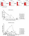
Figure 1. B19-Specific CD8+ T Cells Persist at High Levels for Many Months after Acute Infection
(A) Representative A24 FYT tetramer staining of individual O3\′s PBMCs. Plots are gated on live CD8+ lymphocytes stained directly ex vivo. Percentages shown are those of tetramer-positive CD8+ T cells. Time points indicated refer to the number of months after first symptoms reported. Symptoms in this individual lasted 5 wk. (B) Frequency of B19-specific responses over time for six acutely infected individuals in the Oxford cohort (O1–O5) and five remotely infected individuals (OR1–OR5). In one case, two epitopes were studied. (C) Frequency of B19-specific responses over time for five acutely infected individuals in the Stockholm cohort (S1–S5). In two cases, two epitopes were studied.
Figure 2
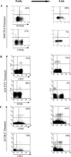
Figure 2. Representative Ex Vivo Phenotyping of B19-Specific CD8+ T Cell Populations over Time
Percentages shown are the frequency of marker-positive cells amongst multimer-positive cells (plots gated on live CD8+ lymphocytes ex vivo). (A) Patient S2 B40 TEA pentamer staining at 4 mo (left) and 21 mo (right), showing perforin and CD62L levels. (B) Patient O3 A24 FYT tetramer staining at 2 mo (left) and 20 mo (right), showing CD27 and CD28 staining. (C) Patient O5 A2 GLC tetramer staining at 4 mo (left) and 18 mo (right), showing CD38 and CD57 staining.
Figure 3
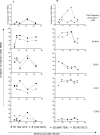
Figure 3. Acutely infected individuals Maintain Activated Mature Effector Cells after Resolution of Acute Infection
Remotely infected individuals show a less mature/activated phenotype but express only low levels of CCR7 and CD62L (Oxford cohort). The y-axis show the frequency of marker-positive cells amongst tetramer-positive CD8+ cells, while the x-axis show the number of months after symptom onset. Data are shown in perforin, CD38, CD57, CD27, CD28, and CCR7: data are derived from six acutely infected individuals in the Oxford cohort (O1–O3, O5, and O6; insufficient cells available for O4) and five remotely infected individuals (OR1–OR5).
Figure 4
Figure 4. Longitudinal Phenotype Analysis of B19-Specific CD8+ T Cell Responses in Acutely Infected Individuals in Stockholm Cohort
(A) Blood from patient S1 was stained with the two tetramers A2 LLH and A2 GLC. (B) Blood from patient S2 was stained with tetramers A2 GLC and the Pro5 B40 TEA pentamer. The top panels show the frequency of tetramer-positive cells over time for the two different responses in each individual (data equivalent to those in Figure 1C). The subsequent panels show the frequency of B19-specific CD8+ cells positive for perforin, CD38, CD57, and CD62L in both patients.
Figure 5
Figure 5. B19-Specific CD8+ T Cells Secrete IFNγ Ex Vivo, Proliferate, and Show Cytolytic Function In Vitro
(A) Left panel shows that PBMCs from acutely infected patient O1 secrete IFNγ ex vivo after 18 h of FYTPLADQF peptide stimulation. Negative control (zero spots) and two peptide-stimulated wells from an ELISpot plate are shown. Numbers represent IFNγ-secreting cells per 250,000 PBMCs. Right panel shows A24 FYT tetramer staining of PBMCs at same time point, displaying the number of tetramer-positive cells expressed as a percentage of CD8+ T cells. (B) Tetramer staining of patient S2\′s PBMCs ex vivo (left) and after short-term TEADVQQWL peptide stimulation in vitro (right). (C) Ex vivo IFNγ ELISpot results for remotely infected individual OR3. Mean and standard deviations of triplicates are shown. Cells were stimulated for 18 h with no peptide, GLCPHCINV, or TEADVQQWL. (D) 51Cr release assay using HLA-A2-restricted GLCPHCINV-specific CTLs from individual OR1. PBMCs were stimulated for 14 d with GLCPHCINV peptide and cytolysis was tested against HLA-A*0201-transfected LBL.721.220 target cells at various effector-to-target-cell (E:T) ratios.

Similar articles

Cited by

References

    1. Gendi NS, Gibson K, Wordsworth BP. Effect of HLA type and hypocomplementaemia on the expression of parvovirus arthritis: one year follow up of an outbreak. Ann Rheum Dis. 1996;55:63–65. - PMC - PubMed
    1. van Elsacker-Niele AM, Kroes AC. Human parvovirus B19: Relevance in internal medicine. Neth J Med. 1999;54:221–230. - PubMed
    1. Heegaard ED, Brown KE. Human parvovirus B19. Clin Microbiol Rev. 2002;15:485–505. - PMC - PubMed
    1. Lundqvist A, Tolfvenstam T, Bostic J, Soderlund M, Broliden K. Clinical and laboratory findings in immunocompetent patients with persistent parvovirus B19 DNA in bone marrow. Scand J Infect Dis. 1999;31:11–16. - PubMed
    1. Tolfvenstam T, Oxenius A, Price DA, Shacklett BL, Spiegel HM, et al. Direct ex vivo measurement of CD8(+) T-lymphocyte responses to human parvovirus B19. J Virol. 2001;75:540–543. - PMC - PubMed

Publication types

MeSH terms

Substances