Review
. 2005 Nov;2(2 Suppl):S9-15.
doi: 10.1016/j.hrthm.2004.09.011.

Role of transmural dispersion of repolarization in the genesis of drug-induced torsades de pointes

Affiliations
Review

Role of transmural dispersion of repolarization in the genesis of drug-induced torsades de pointes

Charles Antzelevitch. Heart Rhythm. 2005 Nov.

Abstract

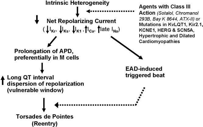
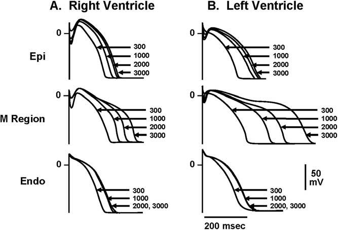
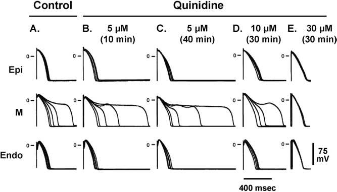
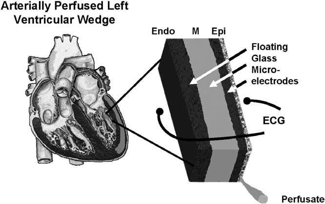
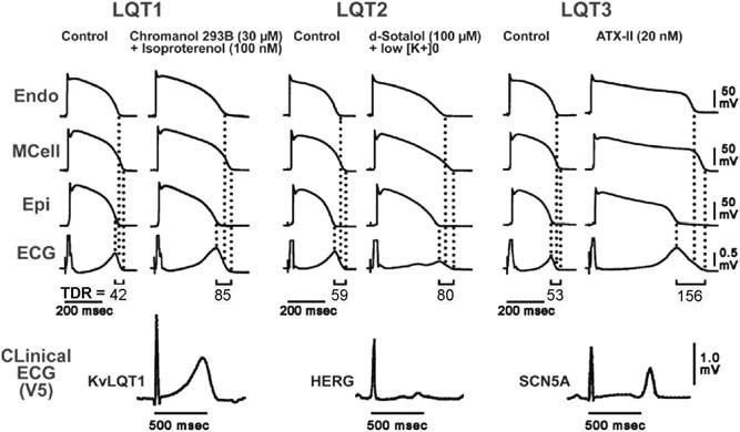
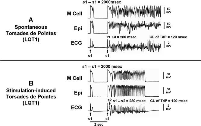
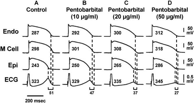
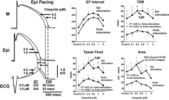
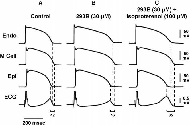
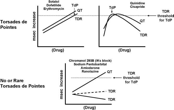
Torsades de pointes (TdP) is a potentially lethal arrhythmia that develops as a consequence of amplification of electrical heterogeneities intrinsic to the ventricular myocardium. These heterogeneities exist because of differences in the time course of repolarization of the three predominant cell types that make up the ventricular myocardium, giving rise to transmural voltage gradients and a dispersion of repolarization responsible for inscription of the ECG T wave. Antiarrhythmic agents with class III actions and/or the various mutations and cardiomyopathies associated with the long QT syndrome reduce net repolarizing current and amplify the intrinsic spatial dispersion of repolarization, thus creating the substrate for the development of reentry. The result is prolongation of the QT interval, abnormal T waves, and development of polymorphic reentrant ventricular tachycardia displaying characteristics of TdP. Prolongation of the QT interval apparently is not the sole determinant of a drug's potential to cause TdP. Agents that do not increase transmural dispersion of repolarization have little or no potential to induce TdP despite any ability to prolong the QT interval. In addition, drugs such as amiodarone and sodium pentobarbital can cause large QT prolongations but, by reducing transmural dispersion of repolarization, may reduce the likelihood of TdP. Arterially perfused wedge preparations of canine left ventricle can be used to explore the role of transmural dispersion of repolarization in the genesis of TdP. The purpose of this article is to review recent advances that have improved our understanding of these mechanisms, particularly the role of transmural dispersion of repolarization, in the genesis of drug-induced TdP and to examine how these advances can guide us toward the development of safer and more effective drugs.


Figures

Figure 1
Figure 1
Mechanism by which transmural dispersion of repolarization and transseptal dispersion of repolarization precipitate torsades de pointes. APD = action potential duration; EAD = early afterdepolarization; M cells = midmyocardial cells.
Figure 2
Figure 2
Cells from the midmyocardial region prolong more than other cell types in response to a slowing of rate, an effect more prominent in the left ventricle than the right ventricle. Endo = endocardial region; Epi = epicardial region; M = midmyocardial. (From Antzelevitch C, Sicouri S, Litovsky SH, Lukas A, Krishnan SC, Di Diego JM, Gintant GA, Liu DW. Heterogeneity within the ventricular wall. Electrophysiology and pharmacology of epicardial, endocardial, and M cells. Circ Res 1991;69:1427–1449, with permission.)
Figure 3
Figure 3
The action potential of M cells is prolonged more than that of epicardial or endocardial cells in the presence agents such as quinidine. Endo = endocardial region; Epi = epicardial region; M = midmyocardial. (From Antzelevitch C, Shimizu W, Yan GX, Sicouri S, Weissenburger J, Nesterenko VV, Burashnikov A, Di Diego J, Saffitz J, Thomas GP. The M cell: its contribution to the ECG and to normal and abnormal electrical function of the heart. J Cardiovasc Electrophysiol 1999;10:1124–1152, with permission from Blackwell Publishing. http://www.blackwell-synergy.com)
Figure 4
Figure 4
Schematic of arterially perfused canine left ventricular wedge preparation. Transmembrane action potentials can be simultaneously recorded from epicardial, M region, endocardial sites or subendocardial Purkinje fibers using three floating microelectrodes. ECG = electrocardiogram.
Figure 5
Figure 5
Transmembrane action potentials and transmural electrocardiograms in control and LQT1, LQT2, and LQT3 models of LQTS (arterially perfused canine left ventricular wedge preparations), and clinical ECG (lead V5) of patients with LQT1 (KvLQT1 defect), LQT2 (HERG defect), and LQT3 (SCN5A defect) syndromes. Isoproterenol plus chromanol 293B, an IKs blocker, d-sotalol plus low K+, and ATX-II, an agent that slows inactivation of late INa, are used to mimic the LQT1, LQT2, and LQT3 syndromes, respectively. ECG = electrocardiogram; Endo = endocardial region; Epi = epicardial region; M = midmyocardial. (From Antzelevitch C, Shimizu W. Cellular mechanisms underlying the long QT syndrome: Curr Opin Cardiol 2002;17:43–51, with permission.)
Figure 6
Figure 6
A: Spontaneous torsades de pointes in an LQT1 model of a wedge preparation. Each trace shows action potentials recorded simultaneously from M and epicardial cells, together with a transmural ECG. B: Stimulation induced torsade de pointes. ECG = electrocardiogram; Epi = epicardial region; M = midmyocardial.
Figure 7
Figure 7
Pentobarbital prolongs the APD of epicardial and endocardial cells and, to a lesser extent, that of the M cell, thus prolonging the QT interval but reducing TDR. The effects of pentobarbital on the QT interval and APD were biphasic. In this study, pentobarbital 10 μg/mL pentobarbital further prolonged APD of epicardial and endocardial cells more than that of the M cell, whereas 20 to 50 μg/mL pentobarbital abbreviated the APD of epicardial and endocardial cells less than that of the M cell, thus abbreviating the QT interval and markedly reducing TDR. Torsades de pointes was never observed, nor could it be induced with programmed electrical stimulation. ECG = electorcardiogram; Endo = endocardial region; Epi = epicardial region; M = midmyocardial; TDR = transmural dispersion of repolarization. (From Shimizu W, McMahon B, Antzelevitch C. Sodium pentobarbital reduces transmural dispersion of repolarization and prevents torsade de pointes in models of acquired and congenital long QT syndrome. J Cardiovasc Electrophysiol 1999;10:154–164, with permission.)
Figure 8
Figure 8
Torsades de pointes (TdP) could be induced with programmed electrical stimulation only at a low concentration of cisapride (0.2 μmol/L), when TDR was maximally prolonged. Moreover, TdP could only be induced during epicardial (but not endocardial) activation of the wedge, which was found to augment TDR. At higher concentrations of cisapride, QT was further prolonged, TDR was diminished, and TdP could no longer be induced. ECG = electrocardiogram; Epi = epicardial region; M = midmyocardial; TdP = torsade de pointes; TDR = transmural dispersion of repolarization. (From Di Diego JM, Belardinelli L, Antzelevitch C. Cisapride-induced transmural dispersion of repolarization and torsades de pointes in the canine left ventricular wedge preparation during epicardial stimulation. Circulation 2003; 108:1027–1033, with permission.)
Figure 9
Figure 9
Transmembrane APs and transmural ECG under control conditions (A), after addition of chromanol 293B 30 μmol/L (B), and after further addition of isoproterenol 100 nmol/L (C). Chromanol 293B prolonged APs of three cell types and QT interval but did not increase TDR; 42–46 ms) or widen the T wave. Isoproterenol in the continued presence of chromanol 293B abbreviated AP of epicardial and endocardial cells but not that of M cells, resulting in an accentuated TDR (85 ms) and broad-based T waves as commonly seen in LQT1 patients. ECG = electrocardiogram; Endo = endocardial region; Epi = epicardial region; M = midmyocardial. (From Shimizu W, Antzelevitch C. Cellular basis for the ECG features of the LQT1 form of the long-QT syndrome: effects of beta-adrenergic agonists and antagonists and sodium channel blockers on transmural dispersion of repolarization and torsade de pointes. Circulation 1998;98:2314 –2322, with permission.)
Figure 10
Figure 10
IKr blockers produce a dose-dependent prolongation of QT that is associated with a dose-dependent accentuation of TDR. When TDR reaches the threshold for reentry, TdP develops. More complex drugs, such as quinidine and cisapride, show a biphasic relationship, with a dose-dependent prolongation of QT until increasing concentrations produce inhibition of inward currents, leading to a reduction of QT. TdP occurs when and if TDR reaches the threshold value for reentry. Other drugs, such as chromanol 293B, pentobarbital, and amiodarone, produce a dose-dependent prolongation of QT but a much smaller change in TDR. Here, threshold values for TdP are rarely reached. TdP is never observed with drugs that prolong QT but produce a dose-dependent reduction of TDR. TdP = torsade de pointes; TDR = transmural dispersion of repolarization.

References

    1. Keating M, Atkinson D, Dunn C, Timothy K, Vincent GM, Leppert M. Linkage of a cardiac arrhythmia, the long-QT syndrome, and the Harvey ras-1 gene. Science. 1991;252:704–706. - PubMed
    1. Ricardo Perez Riera A, Ferreira C, Dubner SJ, Schapachnik E. Andersen syndrome: the newest variant of the hereditary-familial long QT syndrome. Ann Noninvas Electrocardiol. 2004;9:175–179. - PMC - PubMed
    1. Antzelevitch C, Shimizu W. Cellular mechanisms underlying the long QT syndrome. Curr Opin Cardiol. 2002;17:43–51. - PubMed
    1. Antzelevitch C, Sicouri S, Litovsky SH, Lukas A, Krishnan SC, Di Diego JM, Gintant GA, Liu DW. Heterogeneity within the ventricular wall. Electrophysiology and pharmacology of epicardial, endocardial, and M cells. Circ Res. 1991;69:1427–1449. - PubMed
    1. Antzelevitch C, Shimizu W, Yan GX, Sicouri S, Weissenburger J, Nesterenko VV, Burashnikov A, Di Diego J, Saffitz J, Thomas GP. The M cell: its contribution to the ECG and to normal and abnormal electrical function of the heart. J Cardiovasc Electrophysiol. 1999;10:1124–1152. - PubMed

MeSH terms

Substances

LinkOut - more resources