. 2005 Oct 27;33(18):5965-77.
doi: 10.1093/nar/gki905. Print 2005.

Alternative splicing and nonsense-mediated mRNA decay regulate mammalian ribosomal gene expression

Affiliations

Alternative splicing and nonsense-mediated mRNA decay regulate mammalian ribosomal gene expression

Monica Cuccurese et al. Nucleic Acids Res. .

Abstract

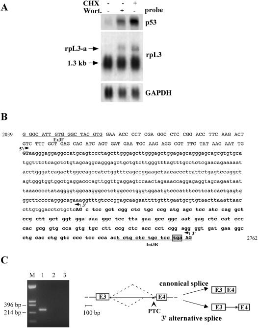
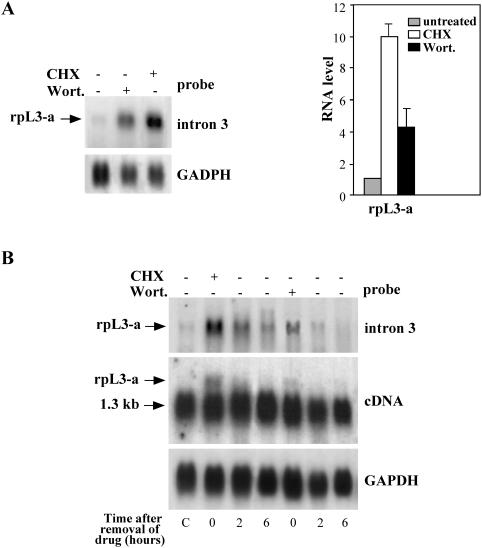
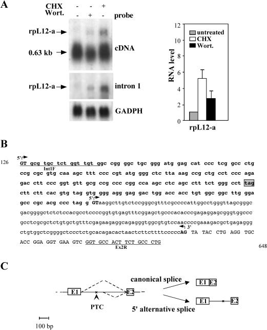
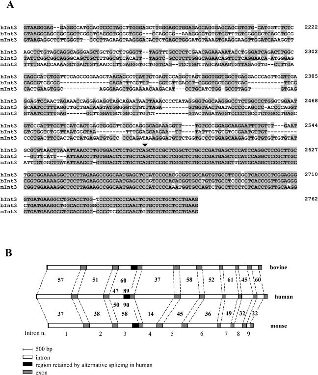
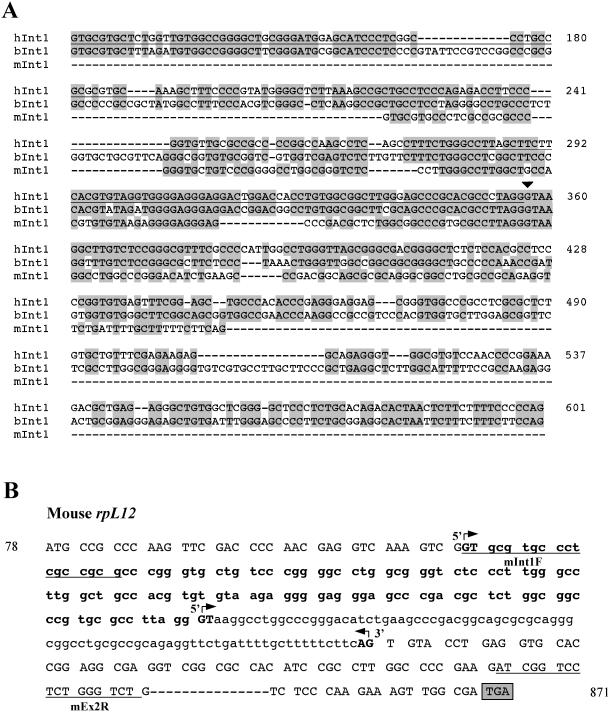
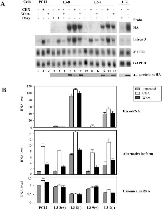
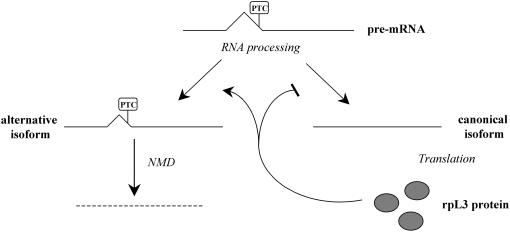
Messenger RNAs containing premature stop codons are generally targeted for degradation through nonsense-mediated mRNA decay (NMD). This mechanism degrades aberrant transcripts derived from mutant genes containing nonsense or frameshift mutations. Wild-type genes also give rise to transcripts targeted by NMD. For example, some wild-type genes give rise to alternatively spliced transcripts that are targeted for decay by NMD. In Caenorhabditis elegans, the ribosomal protein (rp) L12 gene generates a nonsense codon-bearing alternatively spliced transcript that is induced in an autoregulatory manner by the rpL12 protein. By pharmacologically blocking the NMD pathway, we identified alternatively spliced mRNA transcripts derived from the human rpL3 and rpL12 genes that are natural targets of NMD. The deduced protein sequence of these alternatively spliced transcripts suggests that they are unlikely to encode functional ribosomal proteins. Overexpression of rpL3 increased the level of the alternatively spliced rpL3 mRNA and decreased the normally expressed rpL3. This indicates that rpL3 regulates its own production by a negative feedback loop and suggests the possibility that NMD participates in this regulatory loop by degrading the non-functional alternatively spliced transcript.


Figures

Figure 1
Figure 1
Identification of NMD sensitive isoforms of the human rpL3. (A) Analysis of p53 and rpL3 expression in Calu-6 cells after incubation with CHX (4 h) or wortmannin (Wort.) (2 h). Total RNA purified from treated or untreated cells was hybridized with the cDNA specific probes as indicated. The size of canonical mRNA is indicated; rpL3-a indicates the newly identified isoform. The quantity of RNA was normalized using a GAPDH-specific probe. A representative result of three independent experiments is shown. (B) Nucleotide sequence of rpL3 intron 3 with 5′ flanking exonic sequences. Lower case letters refer to intronic nucleotides; upper case letters refer to exonic nucleotides. The intronic sequence retained by alternative splicing is indicated in bold. The primers used for RT–PCR are underlined. Canonical and alternative splice sites (GT/AG), as determined by sequence analysis of RT–PCR products, are shown in bold upper case letters and indicated by 5′ or 3′ arrows. The ORF of exonic and retained intronic sequences is shown. Nucleotides encoding the PTC are boxed and shaded in gray. The nucleotide positions according to the corresponding NCBI/GenBank sequences (accession no.: AL022326) is indicated. (C) Left panel RT–PCR assays of RNA from Calu-6 cells. Amplifications were performed with the following primers: lane 1, Ex3F/Int3R (rpL3-specific); lane 2, reaction was carried out as in lane 1 but omitting the RT step. As negative control, amplification was performed without cDNA (lane 3). The base pairs of HinfII-digested pUC18 (lane M) are indicated on the left. Right panel, schematic drawing of alternative splicing of human rpL3; intron 3 and flanking exons 3 and 4 (E3, E4) are shown.
Figure 2
Figure 2
Expression of alternatively spliced mRNA of the rpL3 gene (rpL3-a mRNA). (A) Left panel: northern blots of total RNA from untreated Calu-6 cells and from the same cells after incubation with either CHX or Wort. The probe used for hybridization is specific for the portion of intron retained by alternative splicing. Right panel: rpL3-a mRNA was quantified by PhosphorImager, normalized to GAPDH levels and expressed in a graph as a ratio to endogenous levels in untreated cells. Numerical values are the average of three independent experiments, reported with SDs. (B) Decay of the drug-stabilized rpL3-a mRNA. Total RNA was isolated from Calu-6 cells before treatment (lane C), after treatment with CHX or with Wort., and 2 and 6 h after drug removal. Northern blot was performed with the indicated probes.
Figure 3
Figure 3
Identification and expression of an alternatively spliced isoform of human rpL12 gene. (A) Left panel: northern blot analysis of rpL12 expression in Calu-6 cells after incubation with CHX (4 h) or Wort. (2 h). Total RNA purified from treated or untreated cells was hybridized with the cDNA specific probes as indicated. The size of canonical mRNA is indicated; rpL12-a indicates the newly identified isoform. The quantity of RNA was normalized using a GAPDH-specific probe. A representative result of three independent experiments is shown. Right panel: rpL12-a mRNA was quantified by PhosphorImager, normalized to GAPDH levels and expressed in a graph as a ratio to endogenous levels in untreated cells. Numerical values are the average of three independent experiments, reported with SDs. (B) Nucleotide sequence of rpL12 intron 1 with 5′ or 3′ flanking exonic sequences. Lower case letters refer to intronic nucleotides; upper case letters refer to exonic nucleotides. The intronic sequence retained by alternative splicing is indicated in bold. The primers used for RT–PCR are underlined. Canonical and alternative splice sites (GT/AG), as determined by sequence analysis of RT–PCR products, are shown in bold upper case letters and indicated by 5′ or 3′ arrows. The ORF of exonic and retained intronic sequences is shown. Nucleotides encoding the PTC are boxed and shaded in gray. The nucleotide positions according to the corresponding NCBI/GenBank sequences (accession no. NT_008470) is indicated. (C) Schematic drawing of alternative splicing of rpL12; the two first exons (E1 and E2) and the first intron are shown.
Figure 4
Figure 4
(A) Sequence comparison of the rpL3 alternatively spliced intron among mammals. Nucleotide sequences of human, bovine and mouse intron 3 (hInt3, bInt3 and mInt3, respectively) are aligned. Conserved residues are shaded in gray; the region retained by alternative splicing is underlined. The numbers on right of sequences indicate the nucleotide positions of the human rpL3 gene according to the corresponding GenBank sequences. Accession nos of sequences (NCBI/GenBank): AJ238851 and NT_039621 for bovine and mouse genes, respectively; for the human gene, refer to the legend to Figure 1 (the arrowhead indicates the alternative splice site). (B) Comparative analysis of rpL3 introns among mammals. The numbers indicate the percent nucleotide identity of human sequences compared to the mouse and bovine genes. For intron 3, the nucleotide identity of the portions retained and excised by alternative splicing is indicated.
Figure 5
Figure 5
Conservation of alternative splicing in the mammalian rpL12 gene. (A) Sequence comparison between human intron 1 (hInt1) and the corresponding bovine (bInt1) and mouse intron (mInt1). The arrowhead indicates the alternative splice site. (B) Nucleotide sequence of mouse rpL12 intron 1 with the 5′ and 3′ flanking exonic sequences. Refer to Figure 4 for alignment; accession nos of sequences (NCBI/GenBank): NT_039206 for mouse gene, NW_619058 for bovine gene; for the human gene, refer to the legend to Figure 3.
Figure 6
Figure 6
Effect of overexpression of r-proteins on the rpL3 splicing pattern. (A) Analysis of rpL3 expression in control cells (PC12) and in clones stably transfected with HA-tagged rpL3 cDNA (L3–8 and L3–9) or with HA-tagged rpL12 cDNA (L12). Northern blot hybridizations of total RNA were performed with the indicated probes. Protein samples purified from the same clones were analyzed by western blotting with an HA-tag specific antibody (αHA). (B) Quantification of mRNAs shown in panel A. The amounts of HA-mRNA are expressed in arbitrary units after normalization to GAPDH mRNA levels. The mRNA levels of endogenous isoforms were normalized to GAPDH and expressed as a ratio to endogenous levels in untreated control cells. (+), addition of doxycycline; (−), removal of doxycycline. The average of three independent experiments is shown, with SDs.
Figure 7
Figure 7
Model for rpL3 feedback regulation. A canonically spliced isoform encodes the rpL3 protein. When present in excess, rpL3 down-regulates canonical splicing and up-regulates the alternative splicing of its own pre-mRNA. This reduces canonical mRNA and increases alternative mRNA. This latter isoform is unproductive and is subsequently eliminated by the NMD pathway.

References

    1. Wagner E., Lykke-Andersen J. mRNA surveillance: the perfect persist. J. Cell Sci. 2002;115:3033–3038. - PubMed
    1. Culbertson M.R., Leeds P.F. Looking at mRNA decay pathways through the window of molecular evolution. Curr. Opin. Genet. Dev. 2003;13:207–214. - PubMed
    1. Gonzalez C.I., Wang W., Peltz S.W. Nonsense-mediated mRNA decay in Saccharomyces cerevisiae: a quality control mechanism that degrades transcripts harboring premature termination codons. Cold Spring Harb. Symp. Quant. Biol. 2001;66:321–328. - PubMed
    1. Ruiz-Echevarria M.J., Czaplinski K., Peltz S.W. Making sense of nonsense in yeast. Trends Biochem. Sci. 1996;21:433–438. - PubMed
    1. Maquat L.E. Nonsense-mediated RNA decay in mammalian cells: A splicing dependent means to down-regulate the levels of mRNAs that prematurely terminate translation. In: Sonenberg N., Hershey J.W.B., Mathews M.B., editors. Translation Control of Gene Expression. NY: Cold Spring Harbor; 2000. pp. 849–868.

Publication types