. 2005 Oct 27;33(18):6000-10.
doi: 10.1093/nar/gki897. Print 2005.

Depletion of TDP 43 overrides the need for exonic and intronic splicing enhancers in the human apoA-II gene

Affiliations

Depletion of TDP 43 overrides the need for exonic and intronic splicing enhancers in the human apoA-II gene

Pablo Arrisi Mercado et al. Nucleic Acids Res. .

Erratum in

Expression of concern in

Abstract

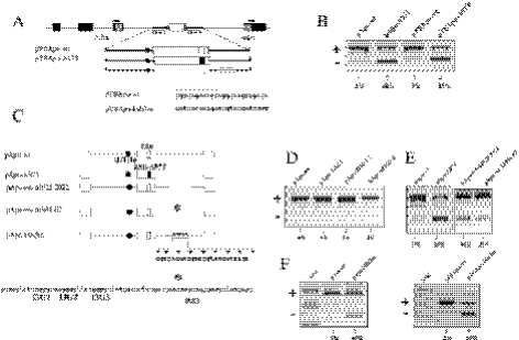
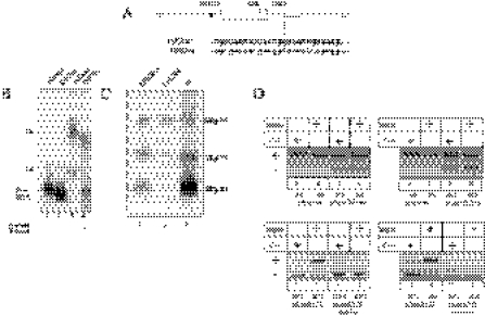
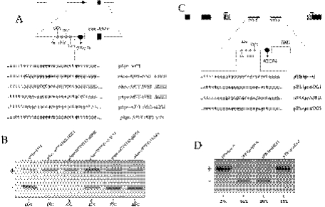
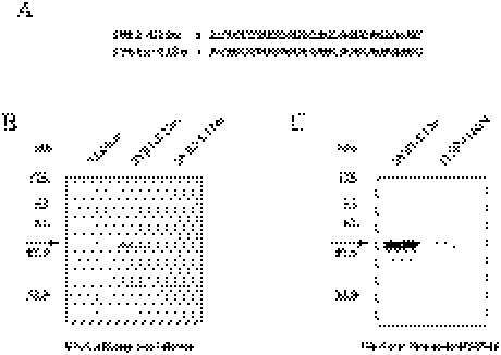
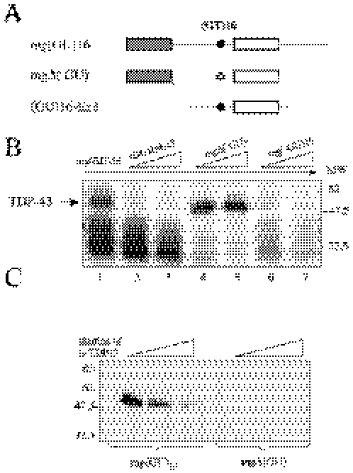
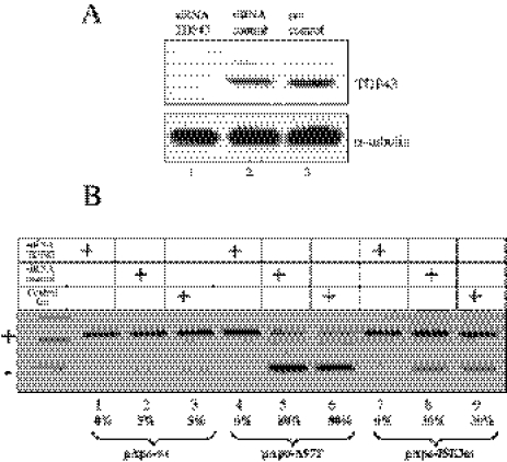
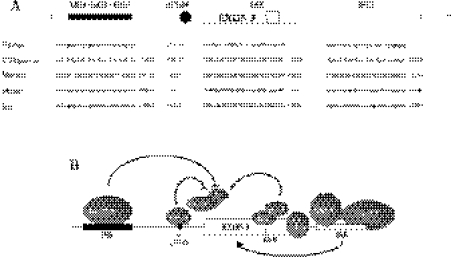
Exon 3 of the human apolipoprotein A-II (apoA-II) gene is efficiently included in the mRNA although its acceptor site is significantly weak because of a peculiar (GU)16 tract instead of a canonical polypyrimidine tract within the intron 2/exon 3 junction. Our previous studies demonstrated that the SR proteins ASF/SF2 and SC35 bind specifically an exonic splicing enhancer (ESE) within exon 3 and promote exon 3 splicing. In the present study, we show that the ESE is necessary only in the proper context. In addition, we have characterized two novel sequences in the flanking introns that modulate apoA-II exon 3 splicing. There is a G-rich element in intron 2 that interacts with hnRNPH1 and inhibits exon 3 splicing. The second is a purine rich region in intron 3 that binds SRp40 and SRp55 and promotes exon 3 inclusion in mRNA. We have also found that the (GU) repeats in the apoA-II context bind the splicing factor TDP-43 and interfere with exon 3 definition. Significantly, blocking of TDP-43 expression by small interfering RNA overrides the need for all the other cis-acting elements making exon 3 inclusion constitutive even in the presence of disrupted exonic and intronic enhancers. Altogether, our results suggest that exonic and intronic enhancers have evolved to balance the negative effects of the two silencers located in intron 2 and hence rescue the constitutive exon 3 inclusion in apoA-II mRNA.


Figures

Figure 1
Figure 1
An ISE sequence is located within the human apoA-II intron 3. (A) Schematic representation of the α-globin/fibronectin reporter system (pTB) used to test the effect of the mutation in the apoA-II exon 3 ESE in a heterologous system α-globin, fibronectin EDB exons and human apoA-II exon 3 are indicated in black, shaded and white boxes, respectively. The black circle indicates the (GT)16 tract. The superimposed arrows indicate the primers used in RT–PCR. The identity of the band is indicated at the left side of the gel. (B) Splicing pattern analysis of the RT–PCR products derived from cellular RNA, stained by ethidium bromide and separated on a 2% agarose gel. Comparison of the effect of the mutation A97T in the apoA-II exon 3 ESE when located in the pApo gene system and in the heterologous α-globin/fibronectin reporter system (pTB). (C) Schematic representation of the constructs carrying deletion and point mutations in the human apoA-II intron 3. White boxes, thin lines and the black circle represent the human apoA-II exons and introns and the (GT)16 tract, respectively. The ESEwt and ESEA97T are indicated as small white and black boxes within the apoA-II exon 3, respectively. Part of the human apoA-II intron 3 sequences. The G runs are in bold. The asterisk indicates a 26 bp poly-purine rich region (underlined) deletion in the apoA-II intron 3. Arrowheads indicate the different point mutations within the polypurine region in intron 3. (D) Splicing pattern analysis by ethidium bromide staining of a 2% agarose gel electrophoresis of the RT–PCR product derived from cellular RNA. The disruption of the G runs in IVS3 was carried out as followed: in the construct pApo-ΔI3G3 ggggctg→gcttatg, in pApo-ΔI3G1–2 gggcaagggg→tcacaagcgc, and in pApo-ΔI3G1–3 gggcaaggggttcagggg→tgtcaagcattcatgcg. (E) Splicing pattern analysis of Hep3B transfected cells with constructs in which partial deletion of the intron 3 were carried out. (D) Schematic representation of the human apoA-II exon 3 and its flanking introns cloned in the α-globin/fibronectin reporter system (pTB). α-globin, fibronectin EDB exons and human apoA-II exon 3 are indicated in black, shaded and white boxes, respectively. The black circle indicates the (GT)16 tract. The small white box within the exon 3 and the white rectangle in the intron 3 represents the ESE and the ISE, respectively. Arrowheads indicate the different point mutations within the polypurine region in intron 3. (F) Comparison of the splicing profiles produced by the mutations in the ISE both in the pApo gene system context and in the pTB reporter minigene. Relative amounts of exon 3 skipping are indicated below the lane numbers. The variability among three different experiments was always <20%.
Figure 2
Figure 2
SRp40 and SRp55 interact with ISE-3 sequence and enhance splicing of exon 3. (A) Schematic representation of the constructs carrying wild-type (ISE3wt) and disrupted (ISE3m) ISE of apoA-II intron 3 used for the EMSA. (B) EMSA of ISE3wt and ISE3m RNAs incubated with HeLa nuclear extract. Complexes were then fractionated on a 4% non-denaturing polyacrylamide gel. The position of RNA–protein complexes (upper complex—Uc and lower complex—Lc) and free RNA are shown. Addition of nuclear extract is also indicated below the lane numbers (C) SDS–PAGE analysis of immunoprecipitation with the specific monoclonal antibody against the phosphorylated RS domain (MAb 1H4) following UV-crosslinking of the labeled RNAs ISE3wt, ISE3m and htot as a positive control (plus) (31). Specific immunoprecipitation of SRp40 (upper panel) and SRp55 (lower panel) is shown only with ISE3wt and htot RNAs. (D) Effects of SRp40 and SRp55 overexpression on apoA-II exon 3 splicing. Relative amounts of exon 3 skipping are indicated below the lane numbers. The variability among three different experiments was always <20%.
Figure 3
Figure 3
A G-rich sequence within the human apoA-II intron 2 influences exon 3 splicing according to context. (A) Schematic representation of the different constructs carrying point mutations in a regulatory sequence within the intron 2 in the pApo gene context. White boxes, thin lines and the black circle represent the human apoA-II exons, introns and the (GT)16 tract, respectively. The ESEA97T is indicated as small black box within the apoA-II exon 3. Sequences within the apoA-II intron 2 changed by site-directed mutagenesis are indicated by small, white and gray circles. Partial sequence of the different constructs carrying point mutations in the intron 2 used for transfection in Hep3B cell line. (B) Agarose gel electrophoresis of RT–PCR products derived from constructs with single G runs or T run disruption in the pApo context. (C) Schematic representation of the different constructs carrying point mutations within the intron 2 of the apoA-II in pTB context. α-globin, fibronectin EDB exons and human apoA-II exon 3 are indicated in black, shaded and white boxes, respectively. Solid black lines, white box and black circle represent the human apoA-II introns, exon 3 and the (GT)16 tract, respectively. The ESEwt is represented by a small white box inside the exon 3. Mutated nucleotides within the apoA-II intron 2 are indicated by small white and gray circles. Partial sequence of the different constructs carrying disrupting point mutations within intron 2. (D) Splicing pattern analysis by ethidium bromide staining of a 2% agarose gel electrophoresis of the RT–PCR products derived from constructs carrying single G run disruption in the pTB context. Relative amounts of exon 3 skipping are indicated below the lane numbers. The variability among three different experiments was always <20%.
Figure 4
Figure 4
The hnRNPH1 binds to G runs within apoA-II intron 2. (A) Sequence of two synthetic RNA oligos carrying the wild-type or the disrupted G runs within the apoA-II intron 2 (IVS2-G12w and IVS2n-G12m, respectively). (B) The Coomassie stained gel of a pull-down assay using adipic dehydrazide beads derivatized with the RNAs following incubation with HeLa nuclear extract. In the lane from the IVS2-G12w-RNA-derivatized beads, the arrow indicates the 58 kDa protein that is absent in the IVS2n-G12m-RNA-derivatized beads. (C) Western blot analysis after the pull-down with the IVS2-G12w and IVS2n-G12m RNA oligos to determine the amount of hnRNPH1 binding.
Figure 5
Figure 5
TDP-43 binds to the (GU) repeats in the apoA-II context. (A) Diagrammatic representation of minigene constructs used for competition experiments and immunoprecipitation with antibodies against TDP-43. Gray and white boxes represent the apoA-II exon 2 and 3, respectively. Thin lines and black circle correspond to the introns and the (GT)16 repeats. Complete deletion of the (GT)16 tract in the construct mgΔ(UG) is indicated by a white cross in the black circle. (B) UV-crosslinking with competition analysis following addition of 5- to 10-fold molar excess of cold (GU)16-Ex3, mgΔ(GU) and mg(GU)16 RNAs to labeled mg(GU)16 RNA in the presence of HeLa nuclear extract. (C) UV-crosslinking plus immunoprecipitation with anti-TDP43 serum. [α-32P]UTP-labeled mg(GU)16 and mgΔ(GU) RNAs were incubated with HeLa nuclear extract before UV-crosslinking. Immunoprecipitation was then carried out with equal amounts of each UV-crosslinked sample using a polyclonal antiserum against TDP-43.
Figure 6
Figure 6
Knock down of TDP-43 causes the rescue of exon 3 inclusion. (A) Upper panel, western blot analysis of the cells treated with the siRNA oligonucleotide against TDP-43 (siRNA TDP43), control siRNA and mock transfected (lanes 1–3, respectively). TDP-43 expression was probed by western blot using a polyclonal antiserum. Lower panel, normalization control western blot with antibody against α-tubulin. (B) Shows splicing pattern analysis by ethidium bromide staining a 2% agarose gel of RT–PCR products after cotransfection of pApo-wt, pApo-A97T and pApo-ISE3m along with either the siRNA TDP43 (lanes 1, 4 and 7) or a control siRNA oligonucleotides (lanes 2, 5 and 8). The transfection of different variants of pApo gene system with no siRNA oligonucleotide was included as a control (lanes 3, 6 and 9).
Figure 7
Figure 7
Evolutionary comparison of the regulatory elements found in human apoA-II exon 3 and its flanking introns in human, macaca, chimpanzee, mouse and rat. (A) Summary of the splicing control elements found in the human apoA-II gene. Black rectangle in intron 2, white square in the exon 3 and white rectangle in intron 3 represent the ISS, ESE and ISE cis-acting elements, respectively. Exon 3 is indicated, thin lines represent introns, and the black circle the (GT)16 tract. The alignment of the different splicing control elements in different species is shown along with the percentage of identity in comparison with human sequences. (B) Cartoon showing a proposed model for the splicing mechanism of the apoA-II exon 3 splicing. TDP-43 and hnRNPH1 prevent the recruitment of U2AF65 to the 3′ splice site of apoA-II intron 2. ASF/SF2 and SC35 interacting with the ESE and SRp40 and SRp55 bound to the ISE might counteract this negative effect by forming a bridge-like structure among components bound to the 5′ and 3′ splice sites permitting the definition of exon 3.

Comment in

References

    1. Smith C.W., Valcarcel J. Alternative pre-mRNA splicing: the logic of combinatorial control. Trends Biochem. Sci. 2000;25:381–388. - PubMed
    1. Black D.L. Mechanisms of alternative pre-messenger RNA splicing. Annu. Rev. Biochem. 2003;72:291–336. - PubMed
    1. Blencowe B.J. Exonic splicing enhancers: mechanism of action, diversity and role in human genetic diseases. Trends Biochem. Sci. 2000;25:106–110. - PubMed
    1. Hovhannisyan R.H., Carstens R.P. A novel intronic cis element, ISE/ISS-3, regulates rat fibroblast growth factor receptor 2 splicing through activation of an upstream exon and repression of a downstream exon containing a noncanonical branch point sequence. Mol. Cell. Biol. 2005;25:250–263. - PMC - PubMed
    1. Arrisi-Mercado P., Romano M., Muro A.F., Baralle F.E. An exonic splicing enhancer offsets the atypical GU-rich 3′ splice site of human apolipoprotein A-II exon 3. J. Biol. Chem. 2004;279:39331–39339. - PubMed

Publication types

MeSH terms

Substances

Grants and funding