. 2005 Nov;79(22):13848-55.
doi: 10.1128/JVI.79.22.13848-13855.2005.

Assembly of severe acute respiratory syndrome coronavirus RNA packaging signal into virus-like particles is nucleocapsid dependent

Affiliations

Assembly of severe acute respiratory syndrome coronavirus RNA packaging signal into virus-like particles is nucleocapsid dependent

Ping-Kun Hsieh et al. J Virol. 2005 Nov.

Abstract

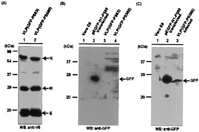
The severe acute respiratory syndrome coronavirus (SARS-CoV) was recently identified as the etiology of SARS. The virus particle consists of four structural proteins: spike (S), small envelope (E), membrane (M), and nucleocapsid (N). Recognition of a specific sequence, termed the packaging signal (PS), by a virus N protein is often the first step in the assembly of viral RNA, but the molecular mechanisms involved in the assembly of SARS-CoV RNA are not clear. In this study, Vero E6 cells were cotransfected with plasmids encoding the four structural proteins of SARS-CoV. This generated virus-like particles (VLPs) of SARS-CoV that can be partially purified on a discontinuous sucrose gradient from the culture medium. The VLPs bearing all four of the structural proteins have a density of about 1.132 g/cm(3). Western blot analysis of the culture medium from transfection experiments revealed that both E and M expressed alone could be released in sedimentable particles and that E and M proteins are likely to form VLPs when they are coexpressed. To examine the assembly of the viral genomic RNA, a plasmid representing the GFP-PS580 cDNA fragment encompassing the viral genomic RNA from nucleotides 19715 to 20294 inserted into the 3' noncoding region of the green fluorescent protein (GFP) gene was constructed and applied to the cotransfection experiments with the four structural proteins. The SARS-CoV VLPs thus produced were designated VLP(GFP-PS580). Expression of GFP was detected in Vero E6 cells infected with the VLP(GFP-PS580), indicating that GFP-PS580 RNA can be assembled into the VLPs. Nevertheless, when Vero E6 cells were infected with VLPs produced in the absence of the viral N protein, no green fluorescence was visualized. These results indicate that N protein has an essential role in the packaging of SARS-CoV RNA. A filter binding assay and competition analysis further demonstrated that the N-terminal and C-terminal regions of the SARS-CoV N protein each contain a binding activity specific to the viral RNA. Deletions that presumably disrupt the structure of the N-terminal domain diminished its RNA-binding activity. The GFP-PS-containing SARS-CoV VLPs are powerful tools for investigating the tissue tropism and pathogenesis of SARS-CoV.


Figures

FIG. 1.
FIG. 1.
Analysis of structural proteins that are assembled into SARS-CoV VLPs. Vero E6 cells previously infected with recombinant vaccinia virus vTF7-3 were cotransfected with plasmids pSec-S(14-1255)-Tag2A, pcDNA-E-V5HisTopo92, pcDNA-M-V5HisTopo158, and pcDNA-N-V5HisTopo56 encoding MycHis-tagged S and V5His-tagged E, M, and N structural proteins, respectively, of SARS-CoV. Four days posttransfection, culture medium was collected, and VLPs were isolated after 20% sucrose cushion centrifugation. Structural proteins assembled into the VLPs were detected by Western blot analysis with mouse polyclonal antibodies generated with 60Co-inactivated SARS-CoV (A) and mouse monoclonal antibody against the V5 epitope of the V5His-tagged structural proteins N, M, and E (B). Lanes 1 and 2 in panel A represent protein lysates of untransfected Vero E6 cells and Vero E6 cells transfected with N-encoding plasmid as negative and positive controls, respectively. Lanes 1 and 2 in panel B represent duplicate experiments.
FIG.2.
FIG.2.
Purification and analysis of SARS-CoV VLPs. (A) E and M proteins could be released from transfected Vero E6 cells in the absence of S and N proteins. To examine the requirements for the formation of SARS-CoV VLPs, Vero E6 cells previously infected with recombinant vaccinia virus vTF7-3 were cotransfected with plasmids encoding the V5His-tagged E and M proteins (lane 2); E, M, and S proteins (lane 3); or E, M, S, and N proteins (lane 4). Culture medium collected 4 days posttransfection was subjected to a 20% sucrose cushion centrifugation and Western blot analysis with the mouse monoclonal antibody against the V5 epitope. Lane 1 represents the nontransfected control. (B) Both E and M proteins could be released into the culture medium when each was expressed alone. Culture medium was collected from Vero E6 cells transfected with plasmids encoding E and M (upper panel), M (middle panel), or E (lower panel). Following a sucrose cushion centrifugation, the pellets were further purified on a discontinuous sucrose gradient as described in Materials and Methods. Fractions were collected and subjected to Western blot analysis. Lanes C represent protein lysates of transfected cells and were used as positive controls. (C) Analysis of different forms of SARS-CoV VLPs. SARS-CoV VLPs formed in the presence of all four structural proteins as demonstrated in panel A (lane 4) were further purified through a discontinuous sucrose gradient. Fractions 4 to 9 were analyzed by Western blot analysis with the V5 epitope-specific antibody for the presence of SARS-CoV structural proteins. Fraction 7 contained the majority of SARS-CoV VLPs, which consisted of all four structural proteins and had a density of around 1.132 g/cm3.
FIG. 3.
FIG. 3.
Secondary structures of the SARS-CoV putative packaging signal PS63 and PS580 RNA. The Mfold program was used to predict the secondary structures of SARS-CoV PScore PS63 RNA encompassing the viral genomic RNA from nt 19888 to 19950 (A) and the PScore-containing PS580 RNA from nt 19715 to 20294 (B). The black rectangle in panel B marks the unique stem-loop structure of the PScore that remains conserved in the PS580 RNA.
FIG. 4.
FIG. 4.
Expression of pEGFP-N1-PS63 and pEGFP-N1-PS580 in Vero E6 cells. Vero E6 cells were transfected individually with pEGFP-N1-PS63 (panels A and B) and pEGFP-N1-PS580 (panels C and D) bearing 63 and 580 nt, respectively, of the putative packaging signals of SARS-CoV inserted into the 3′ noncoding region of the GFP gene. Two days posttransfection, green fluorescence of GFP in the transfected cells was visualized by fluorescence microscopy (panels B and D). Panels A and C show the phase contrast images of the transfected cells.
FIG. 5.
FIG. 5.
Expression of GFP in VLP(GFP-PS580)-infected Vero E6 cells. Plasmid pEGFP-N1-PS63 or pEGFP-N1-PS580 was transfected into Vero E6 cells together with plasmids pSec-S(14-1255)-Tag2A, pcDNA-E-V5HisTopo92, pcDNA-M-V5HisTopo158, and pcDNA-N-V5HisTopo56 that encode a MycHis-tagged S and V5His-tagged E, M, and N structural proteins of SARS-CoV, respectively. Four days posttransfection, VLPs were harvested from the culture medium, partially purified, and used to infect Vero E6 cells to examine the infectivity of the VLPs and the packaging activity of the PS RNAs. The viral proteins that were assembled into the VLPs, VLP(GFP-PS63) and VLP(GFP-PS580), were examined by Western blot analysis with anti-V5 antibody (panel A). Expression of GFP in the VLP(GFP-PS580)-infected cells was examined with anti-GFP antibodies 2 days postinfection (panel C, lane 3). Whether GFP was simultaneously incorporated into the VLPs was also examined (panel B, lanes 3 and 4). Cell lysates prepared from untreated Vero E6 cells (panels B and C, lanes 1) and pEGFP-N1-PS580-transfected cells (panels B and C, lanes 2) were used as negative and positive controls, respectively.
FIG. 6.
FIG. 6.
Packaging of GFP-PS heterologous mRNA into VLPs required sequences beyond the putative 63-nt PScore and was nucleocapsid dependent. Vero E6 cells were infected with either VLP(GFP-PS580) (panels C and D) or VLP(GFP-PS63) (panels E and F). Two days postinfection, green fluorescence in VLP(GFP-PS580)-infected cells was detected by fluorescence microscopy. Panels G and H represent Vero E6 cells that were infected with VLPdN. Untreated Vero E6 cells are shown as negative controls (panels A and B).
FIG.7.
FIG.7.
The RNA-binding activity of the full-length SARS-CoV N protein and its deletion mutants. (A) Schematic representation of the recombinant N proteins and their relative binding activities to PS318 RNA. (B) Coomassie blue staining of the purified N proteins used in the FB assay. BSA and a putative RRM of the eIF3 subunit p116 were used as controls. (C) The FB assay was performed with [α-32P]UTP-labeled PS318 RNA and 40 ng each of the purified N proteins, BSA, and RRM. (D) Specificity of the RNA-binding activity of the N protein subdomains N1 and N2. For determination of the binding specificity of the N1 and N2 subdomains to SARS-CoV PS318 RNA, various amounts of nonlabeled specific competitor (PS318 RNA) and nonspecific competitor (HCV NCR341 RNA) as indicated were mixed with [α-32P]UTP-labeled PS318 RNA before addition of the purified N1 and N2 proteins. Autoradiograms are shown. Relative binding activities (RBA) of the N1 and N2 proteins to the PS318 RNA in the presence of competitors were calculated by normalization of the intensities of signals in each set to that without competitors.

References

    1. Belsham, G. J., J. K. Brangwyn, M. D. Ryan, C. C. Abrams, and A. M. King. 1990. Intracellular expression and processing of foot-and-mouth disease virus capsid precursors using vaccinia virus vectors: influence of the L protease. Virology 176:524-530. - PubMed
    1. Chang, M. F., C. J. Chen, and S. C. Chang. 1994. Mutational analysis of delta antigen: effect on assembly and replication of hepatitis delta virus. J. Virol. 68:646-653. - PMC - PubMed
    1. Chang, M. F., C. Y. Sun, C. J. Chen, and S. C. Chang. 1993. Functional motifs of delta antigen essential for RNA binding and replication of hepatitis delta virus. J. Virol. 67:2529-2536. - PMC - PubMed
    1. Chang, S. C., J. C. Cheng, Y. H. Kou, C. H. Kao, C. H. Chiu, H. Y. Wu, and M. F. Chang. 2000. Roles of the AX4GKS and arginine-rich motifs of hepatitis C virus RNA helicase in ATP- and viral RNA-binding activity. J. Virol. 74:9732-9737. - PMC - PubMed
    1. Chomczynski, P., and N. Sacchi. 1987. Single-step method of RNA isolation by acid guanidinum thiocyanate-phenol-chloroform extraction. Anal. Biochem. 162:156-159. - PubMed

Publication types

LinkOut - more resources