. 2005 Nov;79(22):13856-64.
doi: 10.1128/JVI.79.22.13856-13864.2005.

Kinetic analyses of the surface-transmembrane disulfide bond isomerization-controlled fusion activation pathway in Moloney murine leukemia virus

Affiliations

Kinetic analyses of the surface-transmembrane disulfide bond isomerization-controlled fusion activation pathway in Moloney murine leukemia virus

Michael Wallin et al. J Virol. 2005 Nov.

Abstract

The surface (SU) and transmembrane (TM) subunits of Moloney murine leukemia virus (Mo-MLV) Env are disulfide linked. The linking cysteine in SU is part of a conserved CXXC motif in which the other cysteine carries a free thiol. Recently, we showed that receptor binding activates its free thiol to isomerize the intersubunit disulfide bond into a disulfide within the motif instead (M. Wallin, M. Ekström and H. Garoff, EMBO J. 23:54-65, 2004). This facilitated SU dissociation and activation of TM for membrane fusion. The evidence was mainly based on the finding that alkylation of the CXXC-thiol prevented isomerization. This arrested membrane fusion, but the activity could be rescued by cleaving the intersubunit disulfide bond with dithiothreitol (DTT). Here, we demonstrate directly that receptor binding causes SU-TM disulfide bond isomerization in a subfraction of the viral Envs. The kinetics of the isomerization followed that of virus-cell membrane fusion. Arresting the fusion with lysophosphatidylcholine did not arrest isomerization, suggesting that isomerization precedes the hemifusion stage of fusion. Our earlier finding that native Env was not possible to alkylate but required isomerization induction by receptor binding intimated that alkylation trapped an intermediate form of Env. To further clarify this possibility, we analyzed the kinetics by which the alkylation-sensitive Env was generated during fusion. We found that it followed the fusion kinetics. In contrast, the release of fusion from alkylated, isomerization-blocked virus by DTT reduction of the SU-TM disulfide bond was much faster. These results suggest that the alkylation-sensitive form of Env is a true intermediate in the fusion activation pathway of Env.


Figures
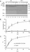
FIG. 1.
FIG. 1.
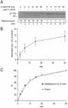
(A) SU-TM disulfide bond isomerization in XC cell-bound Mo-MLV. [35S]Cys-labeled Mo-MLV in culture medium was bound to XC or DF-1 cells for 1 h on ice and incubated in fusion buffer for 0 to 40 min at 37°C. The cultures were lysed in the presence of 20 mM NEM, and viral proteins were captured by immunoprecipitation with polyclonal antibody HE863 for nonreducing SDS-PAGE. The isomerization of the SU-TM disulfide bond was followed by a decrease in SU-TM complexes, increase in free SU, and the appearance of TM. This was observed in virus bound to XC (lanes 1 to 5) but not DF-1 (lanes 7 to 11) cells. Control samples in lanes 6 (XC cell-bound virus) and 12 (DF-1 cell-bound virus) were incubated for 40 min with 1.2 mM M135. Note that free SU does not bind to the DF-1 cells, which are receptor negative. This is in contrast to virus particles, which bind nonspecifically. The figure represents a phosphorimage of the gel. The lower part shows the bottom part of the gel with the TM band at higher contrast. (B) Quantification of isomerization. The degree of isomerization was calculated based on the incubation-induced release of SU from the SU-TM complexes and expressed as a percentage of complete isomerization. In the case of virus bound to DF-1 cells, isomerization was quantified by relating the amount of SU-TM complexes to that of the nonincubated control. (C) Correlation of kinetics of isomerization with that of fusion. The isomerization kinetics was modified from panel B by setting maximal isomerization to 100%. The fusion kinetics was determined by incubating XC cell-bound Mo-MLV at 37°C in fusion buffer for 0 to 40 min and then inactivating the virus by treatment with pH 3 buffer. The cultures were further incubated for 3 h. During this time, virus-fused cells developed into polykaryons, which were used to assess relative fusion efficiencies. Maximal fusion was achieved by 40 min of incubation, which was set to 100%. Standard deviations are indicated.
FIG. 2.
FIG. 2.
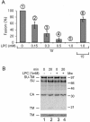
(A) LPC arrests Mo-MLV fusion. Mo-MLV in culture medium was bound to XC cells and incubated in fusion buffer at 37°C for 15 min in the presence of 0 to 1 mM LPC. The virus was then inactivated by pH 3 buffer treatment. Fusion efficiencies (bars 2 to 5) relative to that of a control sample incubated in the absence of LPC (bar 1) were determined as described in the legend to Fig. 1C. Bar 6 shows the relative fusion efficiency of a parallel sample that was first incubated in 1 mM LPC and then washed four times with fusion buffer, incubated for 15 min without LPC, and finally subjected to virus inactivation treatment. Standard deviations are indicated. (B) LPC does not prevent receptor-mediated SU-TM disulfide bond isomerization. [35S]Cys-labeled Mo-MLV was bound to XC- or DF-1 cells and incubated for 0 and 20 min in fusion buffer at 37°C in the presence or absence of 1 mM LPC. Samples were processed for SU-TM disulfide bond isomerization analysis by lysis, immunoprecipitation, and SDS-PAGE as described in the legend to Fig. 1A. Note the similar degrees of SU-TM disulfide bond isomerization in lanes 2 and 4. The figure represents a phosphorimage of the gel. The lower portion shows the TM band at higher contrast.
FIG. 3.
FIG. 3.
(A) Accumulation of alkylated, isomerization-blocked SU-TM complexes during alkylation-inhibited fusion. XC cell-bound [35S]Mo-MLV was incubated at 37°C for 0 to 60 min in fusion buffer containing 1.2 mM M135 and then washed and lysed in the absence (lanes 1 to 6) or presence (lanes 7 to 11) of 1.2 mM M135. SU-TM complexes were captured by immunoprecipitation with the complex-specific MAb 500 and analyzed by nonreducing SDS-PAGE. Note that 10 times more sample has been applied in lanes 1 to 6 than in lanes 7 to 11. The minor amounts of SU observed in most lanes are most likely generated by artificial reduction of the SU-TM disulfide bond during sample preparation for SDS-PAGE (42). (B) Quantification of alkylated, isomerization-blocked SU-TM complexes shown in panel A, lanes 1 to 6. The relative amounts of alkylated complexes at different times of incubation were calculated as percentages of the total amount of viral Env (lanes 7 to 11). (C) Correlation of the kinetics of accumulation of the alkylated, isomerization-blocked SU-TM complexes and that of fusion. The amounts of alkylated complexes at different times of incubation were calculated as percentages of that obtained in the 40-min incubation. The fusion kinetics is from Fig. 1C.
FIG. 4.
FIG. 4.
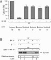
Rapid fusion release of isomerization-arrested state. (A) Mo-MLV was bound to XC cell cultures and then incubated in fusion buffer at 37°C for 15 min in the presence of 1.2 mM M135 to accumulate it at the IAS. The alkylator was washed off, and the cultures were subjected to a second 37°C incubation in fusion buffer with 20 mM DTT for 1 to 15 min. The virus was inactivated by pH 3 treatment, and the DTT-released fusion activity was calculated relative to that of a control sample that was incubated in the absence of alkylator and DTT, as described in the legend to Fig. 1C. (B) [35S]Cys-labeled Mo-MLV was bound to XC cells and then incubated in fusion buffer at 37°C, first for 15 min in the presence of 1.2 mM M135 to induce the IAS and then for 2 min in the presence or absence of 20 mM DTT. The samples were lysed for 50 min at 30°C in the presence or absence of 1.2 mM M135. Intersubunit disulfide bond isomerization-blocked SU-TM complexes were captured by immunoprecipitation with the complex-specific MAb 500 and analyzed by nonreducing SDS-PAGE. Note that 15 times more of samples 3 and 4 than of 1 and 2 were analyzed.
FIG. 5.
FIG. 5.
Hemifusion does not occur at IAS. XC cell-bound Mo-MLV was incubated in fusion buffer at 37°C first for 15 min in the presence of 1.2 mM M135 and then for 1 min in the presence or absence of 0.4 mM CPZ. The effect of CPZ on the fusion reaction of nonarrested virus was tested by incubating cell-bound virus first for 1 min in the presence or absence of 0.4 mM CPZ and then for an additional 15 min without the drug. After the incubations, the virus was inactivated by treatment with pH 3 buffer, and the cultures were processed for polykaryon formation to assess fusion efficiencies. These were expressed as percentages of the control, i.e., the virus fused in the absence of drugs. Note the logarithmic scale.
FIG. 6.
FIG. 6.
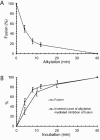
(A) Kinetics of alkylation-induced fusion inhibition. Virus, prebound to XC cells, was subjected to alkylation for 0 to 40 min by incubation at 37°C in fusion buffer containing 1.2 mM M135 and then further incubated in the absence of alkylator for a total time of 40 min. After the cells were washed with virus inactivation buffer, fused cells were allowed to rearrange into polykaryons. Fusion efficiencies relative to that of a control sample incubated for 40 min without alkylator were calculated as described in the legend to Fig. 1C. (B) Correlation between the kinetics of alkylation-induced fusion inhibition and fusion of Mo-MLV. Note that the kinetics of alkylation-induced fusion inhibition is represented as its inverse curve. The fusion kinetics is from Fig. 1C.

Similar articles

Cited by

References

    1. Blumenthal, R., D. P. Sarkar, S. Durell, D. E. Howard, and S. J. Morris. 1996. Dilation of the influenza hemagglutinin fusion pore revealed by the kinetics of individual cell-cell fusion events. J. Cell Biol. 135:63-71. - PMC - PubMed
    1. Brighty, D. W., and S. R. Jassal. 2001. The synthetic peptide P-197 inhibits human T-cell leukemia virus type 1 envelope-mediated syncytium formation by a mechanism that is independent of Hsc70. J. Virol. 75:10472-10478. - PMC - PubMed
    1. Bullough, P. A., F. M. Hughson, J. J. Skehel, and D. C. Wiley. 1994. Structure of influenza haemagglutinin at the pH of membrane fusion. Nature 371:37-43. - PubMed
    1. Carr, C. M., C. Chaudhry, and P. S. Kim. 1997. Influenza hemagglutinin is spring-loaded by a metastable native conformation. Proc. Natl. Acad. Sci. USA 94:14306-14313. - PMC - PubMed
    1. Chan, D. C., D. Fass, J. M. Berger, and P. S. Kim. 1997. Core structure of gp41 from the HIV envelope glycoprotein. Cell 89:263-273. - PubMed

Publication types

LinkOut - more resources