. 2005 Nov;79(22):13882-91.
doi: 10.1128/JVI.79.22.13882-13891.2005.

Viral protein VP4 is a target of human antibodies enhancing coxsackievirus B4- and B3-induced synthesis of alpha interferon

Affiliations

Viral protein VP4 is a target of human antibodies enhancing coxsackievirus B4- and B3-induced synthesis of alpha interferon

Wassim Chehadeh et al. J Virol. 2005 Nov.

Abstract

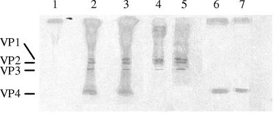
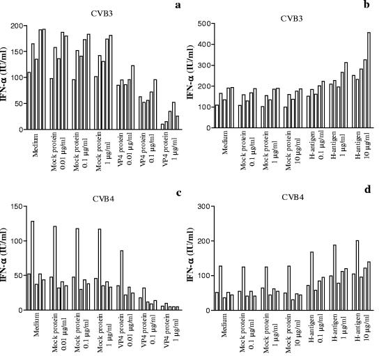
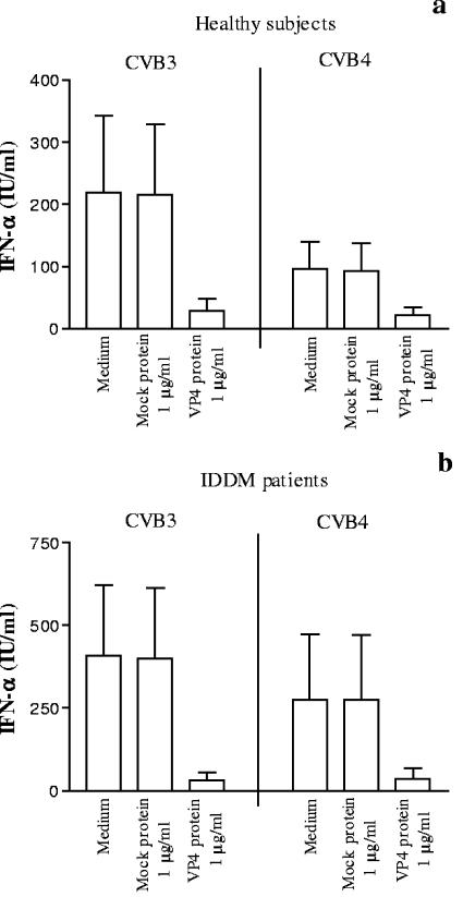
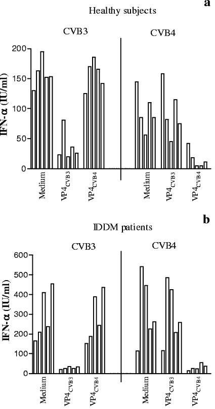
Coxsackievirus B4 (CVB4)-induced production of alpha interferon (IFN-alpha) by peripheral blood mononuclear cells (PBMC) is enhanced in vitro by nonneutralizing anti-CVB4 antibodies from healthy subjects and, to a higher extent, from patients with insulin-dependent diabetes mellitus. In this study, we focused on identification of the viral target of these antibodies in CVB systems. High levels of IFN-alpha were obtained in supernatants of PBMC incubated with CVB4E2 or CVB3 and plasma from healthy subjects and, to a higher extent, from patients. The VP4 capsid proteins dissociated by heating at 56 degrees C from CVB4E2 (VP4(CVB4)) and CVB3 (VP4(CVB3)) but not H antigen preincubated with plasma from healthy subjects or patients inhibited the plasma-dependent enhancement of CVB4E2- and CVB3-induced IFN-alpha synthesis. There was no cross-reaction between VP4(CVB4) and VP4(CVB3) in the inhibiting effect. IFN-alpha levels in culture supernatants showed dose-dependent correlation with anti-VP4 antibodies eluted from plasma specimens using VP4-coated plates. There were higher index values for anti-VP4 antibodies detected by enzyme-linked immunosorbent assay (ELISA) and higher proportions of positive detection in 40 patients than in 40 healthy subjects (80% versus 15% for anti-VP4(CVB4)). There was no relationship between the levels of anti-CVB neutralizing antibodies and the detection of anti-VP4 antibodies by ELISA. The CVB plasma-induced IFN-alpha levels obtained in PBMC cultures in the anti-VP4 antibody-positive groups were significantly higher than those obtained in the anti-VP4 antibody-negative groups regardless of the titers of anti-CVB neutralizing antibodies. These results show that VP4 is the target of antibodies involved in the plasma-dependent enhancement of CVB4E2- and CVB3-induced IFN-alpha synthesis by PBMC.


Figures

FIG. 1.
FIG. 1.
Autoradiogram of CVB capsid polypeptides on 16% sodium dodecyl sulfate-polyacrylamide gel electrophoresis under reducing conditions. Virus polypeptides VP1, VP2, VP3, and VP4 are indicated by lines on the left. Lane 1, mock virus obtained from supernatant of noninfected Hep-2 cells; lanes 2 and 3, 35S-labeled CVB3 and 35S-labeled CVB4E2 from CVB3- and CVB4E2-infected Hep-2 cells, respectively; lanes 4 and 5, H antigen from 35S-labeled CVB3 and 35S-labeled CVB4, respectively, dissociated at 56°C as described in Materials and Methods; lanes 6 and 7, VP4 from 35S-labeled CVB3 and 35S-labeled CVB4E2, respectively, obtained after dissociation.
FIG. 2.
FIG. 2.
Role of VP4 in the plasma-dependent enhancement of CVB-induced IFN-α production by PBMC. Plasma samples obtained from 5 healthy subjects were preincubated for 1 h at 37°C in the absence or presence of the VP4 fraction (a and c) or H antigen (b and d) dissociated from CVB3 or CVB4 (CVB4E2) at various concentrations before the addition of CVB3 or CVB4 (CVB4E2) (MOI, 1) for one additional hour at 37°C. PBMC were then infected with these mixtures. Culture supernatant samples were harvested 48 h postinfection. IFN-α levels in culture supernatants were determined by DELFIA. For each plasma sample, the experiments were performed in duplicate and the culture supernatants obtained for each plasma sample were pooled before the IFN-α assay was performed. Each individual result is presented. Mock protein means protein from mock-infected cells.
FIG. 3.
FIG. 3.
Inhibition of the plasma-dependent enhancement of CVB-induced IFN-α production by PBMC by preincubating plasma with VP4 (1 μg/ml). Plasma samples were obtained from 20 healthy subjects (a) and 20 IDDM patients (b). The procedure is described in the legend to Fig. 2. Means and standard deviations are presented. In the presence of VP4CVB3 protein, the results were 219 ± 123 IU/ml versus 29 ± 20 IU/ml (P < 0.001, n = 20) for healthy subjects and 409 ± 212 IU/ml versus 32 ± 25 IU/ml (P < 0.001, n = 20) for IDDM patients. In the presence of VP4CVB4 protein, the results were 96 ± 44 IU/ml versus 21 ± 13 IU/ml (P < 0.001, n = 20) for healthy subjects and 276 ± 197 IU/ml versus 38 ± 32 IU/ml (P < 0.001, n = 20) for IDDM patients.
FIG. 4.
FIG. 4.
Role of anti-VP4 antibodies in the plasma-dependent enhancement of CVB-induced IFN-α production by PBMC. CVB3 (a) or CVB4 (CVB4E2) (b) was preincubated for 1 h at 37°C with either plasma at optimal dilution obtained from 5 healthy subjects, eluted anti-VP4 antibody fractions at various concentrations obtained from the plasma specimen using VP4-coated plates, anti-VP4-depleted plasma, or control antibodies eluted from mock-infected plates as described in Materials and Methods.
FIG. 5.
FIG. 5.
Absence of cross-reaction between VP4 fractions dissociated from CVB3 or CVB4 (CVB4E2) in the plasma-dependent enhancement of CVB-induced IFN-α production by PBMC. Plasma samples obtained from 5 healthy subjects (a) or 5 IDDM patients (b) were preincubated for 1 h at 37°C in the absence or presence of the VP4 fraction (1 μg/ml) dissociated from CVB3 or CVB4 (CVB4E2) before the addition of CVB3 or CVB4 (CVB4E2) (MOI, 1) for one additional hour at 37°C.
FIG. 6.
FIG. 6.
Individual representation of IFN-α levels in cultures of PBMC obtained from healthy subjects and IDDM patients selected according to the detection of anti-VP4 antibodies. The patients were further segregated according to the titers of neutralizing antibodies (e and f). PBMC were infected with CVB3 (a, b, and c) or CVB4 (CVB4E2) (d, e, and f) preincubated for 1 h at 37°C with plasma obtained from healthy subjects and IDDM patients. The detection of anti-VP4 antibodies was performed by ELISA on plates coated with VP4 dissociated from CVB3 or CVB4 (CVB4E2). Plasma specimens with an index value of anti-VP4 antibodies higher than 1.0 were deemed positive for anti-VP4 antibodies. The horizontal bars represent the means. For anti-VP4CVB3 Ab-positive groups versus anti-VP4CVB3 Ab-negative groups, the results for healthy subjects were 353 ± 87 IU/ml versus 127 ± 42 IU/ml (P < 0.001) and those for IDDM patients were 464 ± 161 IU/ml versus 187 ± 57 IU/ml (P < 0.001) (a and b). For anti-VP4CVB4 Ab-positive groups versus anti-VP4CVB4 Ab-negative groups, the results for healthy subjects were 171 ± 30 IU/ml versus 83 ± 28 IU/ml (P = 0.001) and those for IDDM patients were 305 ± 159 IU/ml versus 104 ± 31 IU/ml (P < 0.001) (d and e). For IDDM patients with low anti-CVB3 NA titers, the results for anti-VP4CVB3-positive antibodies versus anti-VP4CVB3-negative antibodies were 459 ± 154 IU/ml versus 183 ± 55 IU/ml (P < 0.001) (c). For IDDM patients with low anti-CVB4 NA titers, the results for anti-VP4CVB4-positive antibodies versus anti-VP4CVB4-negative antibodies were 388 ± 192 IU/ml versus 104 ± 31 IU/ml (P < 0.001) (f). For IDDM patients with high anti-CVB3 NA titers, the results for anti-VP4CVB3-positive antibodies versus anti-VP4CVB3-negative antibodies were 469 ± 173 IU/ml versus 193 ± 66 IU/ml (P < 0.001) (c). For IDDM patients with anti-VP4CVB4-positive antibodies and low anti-CVB4 NA titers versus high anti-CVB4 NA titers, the results were 388 ± 192 IU/ml versus 241 ± 92 IU/ml (P = 0.01) (f).

Similar articles

Cited by

References

    1. Andreoletti, L., D. Hober, C. Hober-Vandenberghe, S. Belaich, M. C. Vantyghem, J. Lefebvre, and P. Wattre. 1997. Detection of coxsackie B virus RNA sequences in whole blood samples from adult patients at the onset of type I diabetes mellitus. J. Med. Virol. 52:121-127. - PubMed
    1. Bosi, E., R. Minelli, E. Bazzigaluppi, and M. Salvi. 2001. Fulminant autoimmune type 1 diabetes during interferon-alpha therapy: a case of Th1-mediated disease? Diabet. Med. 18:329-332. - PubMed
    1. Boyum, A. 1976. Isolation of lymphocytes, granulocytes and macrophages. Scand. J. Immunol. Suppl. 5:9-15. - PubMed
    1. Brahimi, K., J. L. Perignon, M. Bossus, H. Gras, A. Tartar, and P. Druilhe. 1993. Fast immunopurification of small amounts of specific antibodies on peptides bound to ELISA plates. J. Immunol. Methods 162:69-75. - PubMed
    1. Caggana, M., P. Chan, and A. Ramsingh. 1993. Identification of a single amino acid residue in the capsid protein VP1 of coxsackievirus B4 that determines the virulent phenotype. J. Virol. 67:4797-4803. - PMC - PubMed

Publication types