. 2005 Nov;79(22):13934-42.
doi: 10.1128/JVI.79.22.13934-13942.2005.

A live attenuated vaccine for Lassa fever made by reassortment of Lassa and Mopeia viruses

Affiliations

A live attenuated vaccine for Lassa fever made by reassortment of Lassa and Mopeia viruses

Igor S Lukashevich et al. J Virol. 2005 Nov.

Abstract

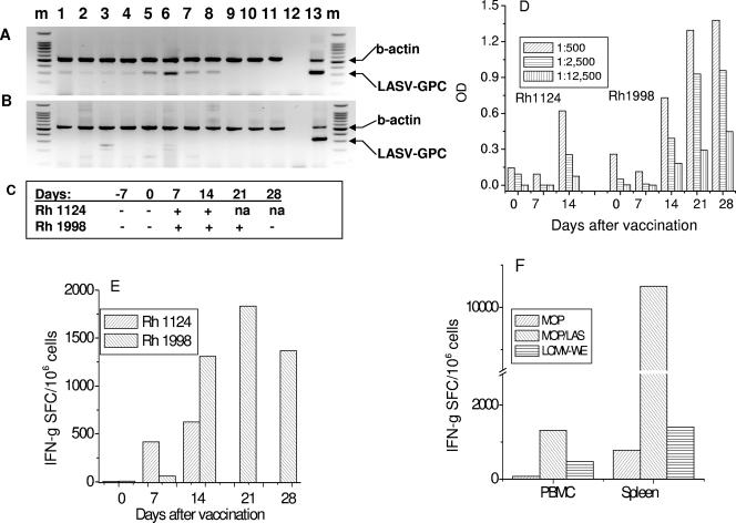
Lassa virus (LASV) and Mopeia virus (MOPV) are closely related Old World arenaviruses that can exchange genomic segments (reassort) during coinfection. Clone ML29, selected from a library of MOPV/LASV (MOP/LAS) reassortants, encodes the major antigens (nucleocapsid and glycoprotein) of LASV and the RNA polymerase and zinc-binding protein of MOPV. Replication of ML29 was attenuated in guinea pigs and nonhuman primates. In murine adoptive-transfer experiments, as little as 150 PFU of ML29 induced protective cell-mediated immunity. All strain 13 guinea pigs vaccinated with clone ML29 survived at least 70 days after LASV challenge without either disease signs or histological lesions. Rhesus macaques inoculated with clone ML29 developed primary virus-specific T cells capable of secreting gamma interferon in response to homologous MOP/LAS and heterologous MOPV and lymphocytic choriomeningitis virus. Detailed examination of two rhesus macaques infected with this MOPV/LAS reassortant revealed no histological lesions or disease signs. Thus, ML29 is a promising attenuated vaccine candidate for Lassa fever.


Figures

FIG. 1.
FIG. 1.
Protective activity of splenocytes from ML29-vaccinated mice. (A) CBA mice were i.p. inoculated with 1,000 PFU of ML29, and at day 7, erythrocyte-free spleen cells were prepared. The recipient mice were i.c. challenged with 1,000 PFU of LASV (time zero), and at different times after challenge, the mice received 30 × 106 immune splenocytes i.v. (B) CBA mice were i.p. inoculated with ML29 at doses varying from 1.5 to 1,500 per animal. At day 7, immune splenocytes were i.v. injected into recipient mice at 3 h after lethal challenge with LASV. (C and D) ML29-immune splenocytes were collected at different time points after vaccination (C) and used at different doses (D) to treat LASV-challenged animals as described above. (E) Tissues from treated and nontreated CBA mice were collected at different time points after challenge and homogenized to prepare 10% (wt/vol) suspensions. Infectious LASV was determined in homogenates by plaque assay. Tissues from four animals were used at each time point. All treated animals survived; nontreated animals died (†) on day 6 after LASV challenge.
FIG. 2.
FIG. 2.
Temperatures, weight measurements, and hemoglobulin levels in plasma of strain 13 guinea pigs. The top three panels show measurements from four mock-infected (PBS-inoculated) and LASV-challenged animals, all of which succumbed (†) within 2 weeks. The lower three panels show measurements from 10 ML29-vaccinated and 10 MOPV-vaccinated guinea pigs, all of which survived LASV challenge for at least 70 days. The error bars indicate standard deviations from the data points.
FIG. 3.
FIG. 3.
Liver enzymes in ML29-vaccinated animals challenged with LASV. ALT, AST, and AlkPh were monitored in control and vaccinated animals as a sign of LF. The top left panel shows the elevation of these markers within 8 to 12 days after LASV challenge for the animals that were not vaccinated and died from LF. The remaining three panels show levels of liver enzymes in 10 guinea pigs vaccinated with ML29 (MOP/LAS) compared to 10 guinea pigs vaccinated with MOPV. The error bars indicate standard deviations from the data points.
FIG. 4.
FIG. 4.
Histology of lung (A to D) and liver (E to H), hematoxylin and eosin staining. (A and E) LASV-infected animals. (B and F) MOP/LAS-vaccinated and LASV-challenged guinea pigs. (C and G) MOPV-vaccinated and LASV-challenged animals. (D and H) Tissues of normal guinea pigs.
FIG. 5.
FIG. 5.
Infection of rhesus macaques with ML29 reassortant. (A and B) Detection of ML29 sequences in tissues. Rhesus macaques were vaccinated with ML29 and sacrificed on day 14 (Rh 1124) (A) and day 28 (Rh 1998) (B). RNA samples were isolated, converted into cDNA, and amplified with LASV 36E2 and 80F2 primers (8). Lanes: 1, lung; 2, stomach; 3, liver; 4, kidney; 5, MLN; 6, spleen; 7, ileum; 8, heart; 9, cerebrum; 10, cerebellum; 11, hippocampus; 12, negative PCR control; 13, positive PCR control (RNA from LASV-infected Vero E6 cells); m, 100-bp DNA ladder. The arrows indicate the positions of β-actin (540 bp) and LASV (335 bp) amplicons. (C) Viremia in vaccinated animals detected by RT-PCR. +, RT-PCR positive; −, RT-PCR negative; n/a, not applicable. (D) IgG ELISA. Serum samples collected weekly were assayed with ML29 antigen as described in Materials and Methods. (E) T cells from peripheral blood secreting IFN-γ in response to in vitro stimulation with ML29. (F) IFN-γ-secreting T lymphocytes in spleen (28 days after vaccination) in response to specific ML29 and closely related antigens, MOPV and LCMV WE.

References

    1. Anonymous. 2003. Influenza virus vaccine live intranasal-MedImmune vaccines: CAIV-T, influenza vaccine live intranasal. Drugs R&D 4:312-319. - PubMed
    1. Auperin, D. D., D. R. Sasso, and J. B. McCormick. 1986. Nucleotide sequence of the glycoprotein gene and intergenic region of the Lassa virus S genome RNA. Virology 154:155-167. - PubMed
    1. Borio, L., T. Inglesby, C. J. Peters, A. L. Schmaljohn, J. M. Hughes, P. B. Jahrling, T. Ksiazek, K. M. Johnson, A. Meyerhoff, T. O'Toole, M. S. Ascher, J. Bartlett, J. G. Breman, E. M. Eitzen, Jr., M. Hamburg, J. Hauer, D. A. Henderson, R. T. Johnson, G. Kwik, M. Layton, S. Lillibridge, G. J. Nabel, M. T. Osterholm, T. M. Perl, P. Russell, K. Tonat, and the Working Group on Civilian Biodefense. 2002. Hemorrhagic fever viruses as biological weapons: medical and public health management. JAMA 287:2391-2405. - PubMed
    1. Bowen, M., P. Rollin, T. Ksiazek, H. Hustad, D. Bausch, A. Demby, M. Bajani, C. Peters, and S. Nichol. 2000. Genetic diversity among Lassa virus strains. J. Virol. 74:6992-7004. - PMC - PubMed
    1. Chandler, L. J., G. Hogge, M. Endres, D. R. Jacoby, N. Nathanson, and B. J. Beaty. 1991. Reassortment of La Crosse and Tahyna bunyaviruses in Aedes triseriatus mosquitoes. Virus Res. 20:181-191. - PubMed

Publication types

MeSH terms

LinkOut - more resources