. 2005 Nov;79(22):14079-87.
doi: 10.1128/JVI.79.22.14079-14087.2005.

Laser-capture microdissection: refining estimates of the quantity and distribution of latent herpes simplex virus 1 and varicella-zoster virus DNA in human trigeminal Ganglia at the single-cell level

Affiliations

Laser-capture microdissection: refining estimates of the quantity and distribution of latent herpes simplex virus 1 and varicella-zoster virus DNA in human trigeminal Ganglia at the single-cell level

Kening Wang et al. J Virol. 2005 Nov.

Abstract

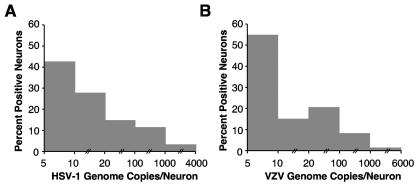
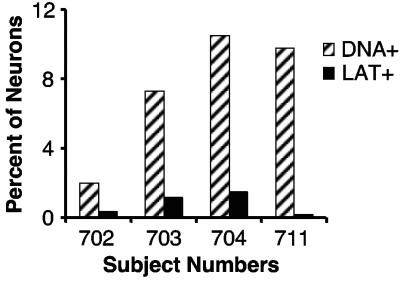
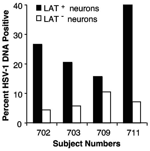
There remains uncertainty and some controversy about the percentages and types of cells in human sensory nerve ganglia that harbor latent herpes simplex virus 1 (HSV-1) and varicella-zoster virus (VZV) DNA. We developed and validated laser-capture microdissection and real-time PCR (LCM/PCR) assays for the presence and copy numbers of HSV-1 gG and VZV gene 62 sequences in single cells recovered from sections of human trigeminal ganglia (TG) obtained at autopsy. Among 970 individual sensory neurons from five subjects, 2.0 to 10.5% were positive for HSV-1 DNA, with a median of 11.3 copies/positive cell, compared with 0.2 to 1.5% of neurons found to be positive by in situ hybridization (ISH) for HSV-1 latency-associated transcripts (LAT), the classical surrogate marker for HSV latency. This indicates a more pervasive latent HSV-1 infection of human TG neurons than originally thought. Combined ISH/LCM/PCR assays revealed that the majority of the latently infected neurons do not accumulate LAT to detectable levels. We detected VZV DNA in 1.0 to 6.9% of individual neurons from 10 subjects. Of the total 1,722 neurons tested, 4.1% were VZV DNA positive, with a median of 6.9 viral genomes/positive cell. After removal by LCM of all visible neurons on a slide, all surrounding nonneuronal cells were harvested and assayed: 21 copies of HSV-1 DNA were detected in approximately 5,200 nonneuronal cells, while nine VZV genomes were detected in approximately 14,200 nonneuronal cells. These data indicate that both HSV-1 and VZV DNAs persist in human TG primarily, if not exclusively, in a moderate percentage of neuronal cells.


Figures

FIG. 1.
FIG. 1.
Schematic representation of LCM. An infrared laser is integrated into a PixCell II microscope. A cap containing a transparent, thermoplastic membrane is placed directly on the surface of a tissue section on a glass slide. A laser pulse heats and activates a defined area of the membrane, causing it to adhere to (A) and bind the targeted portion of the tissue (B). DNA is then extracted from the captured cells for PCR analyses.
FIG. 2.
FIG. 2.
LCM of neurons and satellite cells from human trigeminal ganglion sections. (A and B) Photomicrographs (100×) of a ganglion section before and after dissection of single neurons, respectively. The arrows indicate the neurons in which nuclei were clearly visible (A) and those which were microdissected completely (B). It is evident in panel B that nonneuronal, satellite cells surrounding the targeted neurons were retained in the tissue section. (C) Several captured neurons on a cap. (D) Photomicrograph of a tissue section in which neurons had been removed from the circled clusters but the satellite cells surrounding those neurons were retained in the section. (E) The same section as in panel D after the satellite cells in the circled clusters had been removed by LCM. (F) Some captured satellite cells on a cap.
FIG. 3.
FIG. 3.
HSV-1 and VZV genome copy numbers in individual PCR-positive neurons. A total of 970 single neurons captured from sections from five study subjects were tested for HSV-1 gG, of which 61 were PCR positive, with genome copy numbers/cell ranging from 5 (the lower limit of detection of the PCR assay) to 3,955 and with a median of 11.3 copies/positive cell. A total of 71% of all positive neurons contained 20 or fewer copies of HSV-1 DNA (A). The VZV genome copy number detected in single neurons ranged from 2.6 copies (the lower limit of detection of the assay being 5 copies of the diploid gene ORF62, or 2.5 genomes) to 5,773 copies, with a median of 6.9 copies per positive cell (B). The VZV genome copies per positive cell were ≤20 in 70% of all positive neurons.
FIG. 4.
FIG. 4.
LCM of LAT-positive and LAT-negative neurons. Human TG sections were subjected to in situ hybridization for HSV-1 LAT. The hybridization signals were visualized microscopically and photographed while sections were covered with xylene. (A) ISH section covered with xylene, in which five LAT-positive neurons could be identified (200×). (B and C) The section was then air dried (B), and each LAT-positive neuron was harvested by LCM (C). The LAT-negative neurons were then harvested from these sections after all clearly LAT-positive neurons were removed.
FIG. 5.
FIG. 5.
More neurons contain HSV-1 DNA than are found to contain LAT. DNA extracted from LCM caps bearing single neurons was subjected to real-time PCR for HSV-1 gG sequences. Adjacent sections were subjected to ISH for HSV-1 LAT. For sections of all four subjects studied, the percentages (2.0, 7.3, 10.5, and 9.8) of neurons that were PCR positive for HSV-1 DNA (DNA+) were higher than the percentages (0.4, 1.2, 1.5, and 0.2) of cells that were LAT positive (LAT+).
FIG. 6.
FIG. 6.
HSV-1 DNA is detected in LAT-negative neurons. Single LAT-positive or LAT-negative neurons dissected from human TG sections, as shown in Fig. 4, of subjects 702, 703, 709, and 711 were subjected to real-time PCR for HSV-1 gG sequences. HSV-1 DNA was detected in 4, 6, 11, and 7% of LAT-negative neurons, respectively. In LAT-positive neurons, the percentages of viral DNA-positive cells were higher, at 27, 21, 16, and 40%, respectively.

Similar articles

Cited by

References

    1. Ashley, R. L., J. Militoni, F. Lee, A. Nahmias, and L. Corey. 1988. Comparison of Western blot (immunoblot) and glycoprotein G-specific immunodot enzyme assay for detecting antibodies to herpes simplex virus types 1 and 2 in human sera. J. Clin. Microbiol. 26:662-667. - PMC - PubMed
    1. Bloom, D. C., G. B. Devi-Rao, J. M. Hill, J. G. Stevens, and E. K. Wagner. 1994. Molecular analysis of herpes simplex virus type 1 during epinephrine-induced reactivation of latently infected rabbits in vivo. J. Virol. 68: 1283-1292. - PMC - PubMed
    1. Bloom, D. C., J. M. Hill, G. Devi-Rao, E. K. Wagner, L. T. Feldmen, and J. G. Stevens. 1996. A 348-base-pair region in the latency-associated transcript facilitates herpes simplex virus type 1 reactivation. J. Virol. 70:2449-2459. - PMC - PubMed
    1. Burke, R. L., K. Hartog, K. D. Croen, and J. M. Ostrove. 1991. Detection and characterization of latent HSV RNA by in situ and Northern blot hybridization in guinea pigs. Virology 181:793-797. - PubMed
    1. Cai, G. Y., L. I. Pizer, and M. J. Levin. 2002. Fractionation of neurons and satellite cells from human sensory ganglia in order to study herpesvirus latency. J. Virol. Methods 104:21-32. - PubMed

Publication types

MeSH terms