. 2005 Nov;79(22):14102-11.
doi: 10.1128/JVI.79.22.14102-14111.2005.

Epstein-Barr virus mRNA export factor EB2 is essential for intranuclear capsid assembly and production of gp350

Affiliations

Epstein-Barr virus mRNA export factor EB2 is essential for intranuclear capsid assembly and production of gp350

Julien Batisse et al. J Virol. 2005 Nov.

Abstract

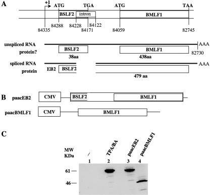
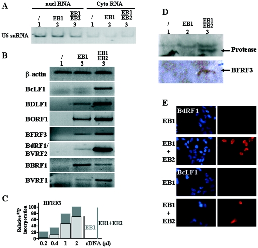
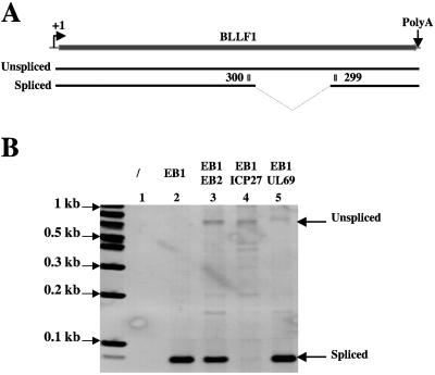
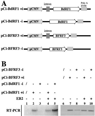
Most human herpesviruses, including Epstein-Barr virus (EBV), express a protein which functions primarily as an mRNA export factor. Previously, we deleted the gene for the Epstein-Barr virus mRNA export factor EB2 from the EBV genome and then introduced the mutated genome into 293 cells. Using a transcomplementation assay in which ectopic expression of the transcription factor EB1/ZEBRA was sufficient to induce the EBV productive cycle, we showed that Ori-Lyt-dependent replication of the EBV DNA occurs in the absence of EB2, indicating that EB2 is not essential for the expression and export of early mRNAs. However, in the absence of EB2, no infectious viral particles are produced (H. Gruffat, J. Batisse, D. Pich, B. Neuhierl, E. Manet, W. Hammerschmidt, and A. Sergeant, J. Virol. 76:9635-9644, 2002). In this report, we now show that EB2 is essential for the nuclear export of most, but not all, late mRNAs produced from intronless genes that translate into proteins involved in intranuclear capsid assembly and maturation. As a consequence, we show that EB2 is essential for the proper assembly of intranuclear capsids. Interestingly, the late BLLF1 gene contains an intron, and both unspliced and spliced mRNAs must be exported to the cytoplasm to be translated into gp350 and gp220, respectively (M. Hummel, D. A. Thorley-Lawson, and E. Kieff, J. Virol. 49:413-417, 1984). Our results also demonstrate that although BLLF1 spliced mRNAs are exported to the cytoplasm independently of EB2, EB2 is essential for the nuclear export of unspliced BLLF1 mRNA. In the same assay, herpes simplex virus 1 ICP27 completely inhibited the nuclear export of BLLF1 spliced mRNAs whereas unspliced BLLF1 mRNAs were exported, confirming that in a physiological assay, ICP27 inhibits splicing.


Figures

FIG. 1.
FIG. 1.
EB2 is the product of the BSLF2/BMLF1 gene. (A) Schematic structure of the BSLF2 and BMLF1 open reading frames on the EBV genome. The relative positions of the initiation codons (ATG), stop codons, the borders of the intron, and the putative ORFs are given according to the sequence of the B95-8 viral strain. (B) Schematic representation of the expression vectors used. The cDNA sequence from the BSLF2/BMLF1 gene was inserted in the paac vector to generate the paacEB2 vector. paacBMLF1 contains the BMLF1 ORF. CMV, cytomegalovirus promoter. (C) Western blot analysis of Raji cells transfected with the expression vector paacEB2 (lane 3) or paacBMLF1 (lane 4) or induced into the productive cycle by treatment with TPA/BA (lane 2). A rabbit polyclonal serum directed against the EB2 protein was used.
FIG. 2.
FIG. 2.
Packaging of the EBV DNA in 293BMLF1-ko cells. 293BMLF1-ko cells were induced into the productive cycle by transfection with an EB1 expression vector and transcomplemented or not transcomplemented with an EB2 expression vector as indicated above the lanes. Total (/) and DNase-resistant (DNase I) DNAs were prepared 48 h after transfection. Samples were cleaved with BamHI, and the fragments were resolved by electrophoresis through a 0.8% agarose gel, transferred to a nylon membrane, and hybridized to 32P-labeled BRRF1 DNA. The numbers below the lanes containing total DNA indicate the ratios of newly replicated viral DNA quantified after PhosphorImager exposure of the membrane. As a control for the amount of DNA loaded on the gel, the Southern blot was also incubated with a β-globin probe.
FIG. 3.
FIG. 3.
The EB2 protein induces the cytoplasmic accumulation of specific mRNA encoding EBV capsid protein. (A) Control of the purity of our cytoplasmic (Cyto) RNA fraction: RNA from 293BMLF1-ko cells transiently transfected with a control vector or expression plasmids encoding proteins as indicated at the top of the figure were submitted to RT-PCR analysis using specific primers to detect the U6 snRNA. nucl, nuclear. (B) RNA from 293BMLF1-ko cells transiently transfected as indicated at the top of the figure were submitted to RT-PCR analysis using specific primers to detect the β-actin cellular mRNA or EBV late mRNA encoding BcLF1, BDLF1, BORF1, BFRF3, BdRF1, BBRF1, and BVRF1. The PCR products were loaded on a 2% agarose gel and visualized by ethidium bromide staining. (C) Example from the results of our semiquantitative experiments done using the BFRF3 mRNA. Each PCR (25 cycles) was done with various amounts of RT product and in the presence of [α-32P]dCTP. The gel was quantified after PhosphorImager exposure. (D) Western blot analysis of the 293BMLF1-ko cells transiently transfected with a control vector or expression plasmids encoding proteins as indicated at the top of the figure. The membranes were incubated with either an anti-protease or an anti-BFRF3 monoclonal antibody. (E) Expression of the BdRF1 and BcLF1 proteins was monitored by immunofluorescence. The 293BMLF1-ko cells were transiently transfected with expression plasmids encoding proteins as indicated on the left of the figure. After the cells were labeled with the appropriate antibody, they were stained with bisBENZIMIDE to label their nuclei and observed on a UV fluorescence microscope. The nuclei stained blue; the BdRF1 and BcLF1 proteins stained red.
FIG. 4.
FIG. 4.
Cytoplasmic accumulation of late BALF4 mRNA is EB2 independent. (A) RNA from 293BMLF1-ko cells transiently transfected with a control vector or expression plasmids encoding proteins as indicated at the top of the figure were submitted to RT-PCR analysis using specific primers to detect the BALF4 mRNA. The PCR (25 cycles) was done with various amounts of RT product and in the presence of [α-32P]dCTP. The PCR products were loaded on a 6% polyacrylamide gel and visualized by autoradiography. (B) Expression of the BALF4 protein (gp110) was monitored by immunofluorescence. The 293BMLF1-ko cells were transiently transfected with expression plasmids encoding proteins as indicated on the left of the figure. After the cells were labeled with the anti-BALF4 antibody, they were observed on a UV fluorescence microscope. The BALF4 protein stained red.
FIG. 5.
FIG. 5.
Expression of the late BLLF1 gene. (A) Schematic representation of the BLLF1 gene. +1, mRNA initiation site; PolyA, polyadenylation signal. From the BLLF1 gene, by alternative splicing, two mRNAs are synthesized encoding, respectively, the gp350 and gp220 proteins. 299 and 300 are the primers used for the PCR. (B) RNA from 293BMLF1-ko cells transiently transfected with a control vector or expression plasmids encoding proteins as indicated at the top of the panel were submitted to RT-PCR analysis using specific primers (299 and 300) to detect the two BLLF1 mRNAs. The PCR products were loaded on a 2% agarose gel and visualized by ethidium bromide staining.
FIG. 6.
FIG. 6.
The EB2-dependent cytoplasmic accumulation of intronless EBV mRNAs can be reproduced in a transient expression assay with nonreplicating plasmids. (A) Schematic representation of the reporter plasmids used. The BFRF3 or BdRF1 ORF was cloned under the control of the CMV promoter into the pCI vector containing or not containing an artificial intron. (B) RNA from HeLa cells transiently transfected with a control vector or expression plasmids encoding proteins as indicated at the top of the panel were submitted to RT-PCR analysis using specific primers to detect the BFRF3 or the BdRF1 mRNA. The PCR products were loaded on a 2% agarose gel and visualized by ethidium bromide staining.

Similar articles

Cited by

References

    1. Amon, W., and P. J. Farrell. 2005. Reactivation of Epstein-Barr virus from latency. Rev. Med. Virol. 15:149-156. - PubMed
    1. Babcock, G. J., L. L. Decker, M. Volk, and D. A. Thorley-Lawson. 1998. EBV persistence in memory B cells in vivo. Immunity 9:395-404. - PubMed
    1. Boyer, J. L., S. Swaminathan, and S. J. Silverstein. 2002. The Epstein-Barr virus SM protein is functionally similar to ICP27 from herpes simplex virus in viral infections. J. Virol. 76:9420-9433. - PMC - PubMed
    1. Buisson, M., F. Hans, I. Kusters, N. Duran, and A. Sergeant. 1999. The C-terminal region but not the Arg-X-Pro repeat of Epstein-Barr virus protein EB2 is required for its effect on RNA splicing and transport. J. Virol. 73:4090-4100. - PMC - PubMed
    1. Buisson, M., E. Manet, M. C. Biemont, H. Gruffat, B. Durand, and A. Sergeant. 1989. The Epstein-Barr virus (EBV) early protein EB2 is a posttranscriptional activator expressed under the control of EBV transcription factors EB1 and R. J. Virol. 63:5276-5284. - PMC - PubMed

Publication types