. 2005 Nov;79(22):14339-45.
doi: 10.1128/JVI.79.22.14339-14345.2005.

PrPTSE distribution in a primate model of variant, sporadic, and iatrogenic Creutzfeldt-Jakob disease

Affiliations

PrPTSE distribution in a primate model of variant, sporadic, and iatrogenic Creutzfeldt-Jakob disease

Christian Herzog et al. J Virol. 2005 Nov.

Abstract

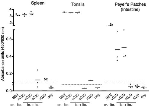
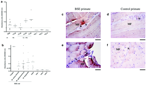
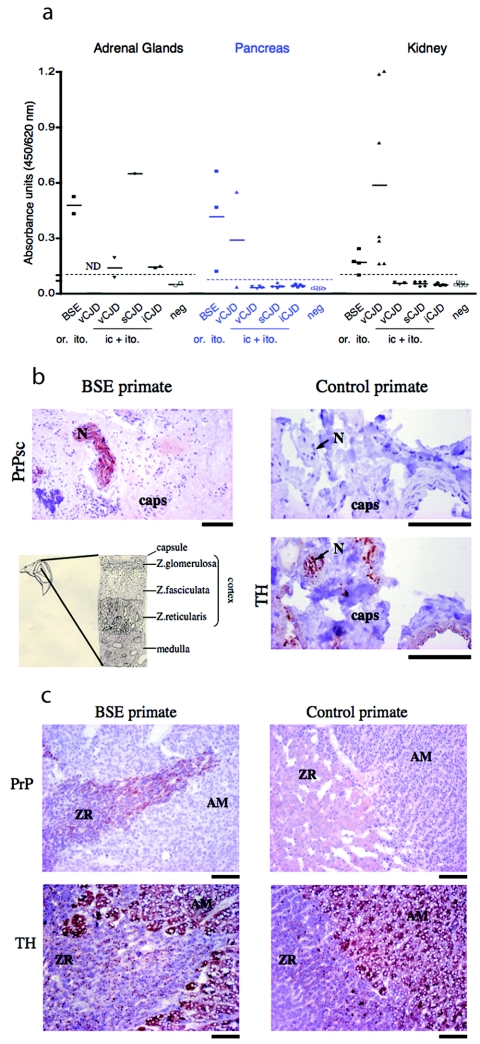
Human prion diseases, such as Creutzfeldt-Jakob disease (CJD), are neurodegenerative and fatal. Sporadic CJD (sCJD) can be transmitted between humans through medical procedures involving highly infected organs, such as the central nervous system. However, in variant CJD (vCJD), which is due to human contamination with the bovine spongiform encephalopathy (BSE) agent, lymphoreticular tissue also harbors the transmissible spongiform encephalopathy-associated prion protein (PrP(TSE)), which poses a particularly acute risk for iatrogenic transmission. Two blood transfusion-related cases are already documented. In addition, the recent observation of PrP(TSE) in spleen and muscle in sCJD raised the possibility that peripheral PrP(TSE) is not limited to vCJD cases. We aimed to clarify the peripheral pathogenesis of human TSEs by using a nonhuman primate model which mimics human diseases. A highly sensitive enzyme-linked immunosorbent assay was adapted to the detection of extraneural PrP(TSE). We show that affected organs can be divided into two groups. The first is peripheral organs accumulating large amounts of PrP(TSE), which represent a high risk of iatrogenic transmission. This category comprises only lymphoreticular organs in the vCJD/BSE model. The second is organs with small amounts of PrP(TSE) associated with nervous structures. These are the muscles, adrenal glands, and enteric nervous system in the sporadic, iatrogenic, and variant CJD models. In contrast to the first set of organs, this low level of tissue contamination is not strain restricted and seems to be linked to secondary centrifugal spread of the agent through nerves. It might represent a risk for iatrogenic transmission, formerly underestimated despite previous reports of low rates of transmission from peripheral organs of humans to nonhuman primates (5, 10). This study provides an additional experimental basis for the classification of human organs into different risk categories and a rational re-evaluation of current risk management measures.


Figures

FIG. 1.
FIG. 1.
Sensitivity of PrPTSE detection in different peripheral organs. Tissue samples were spiked with different amounts of infectious brain homogenate to determine the detection level. Linear regression analysis was performed to generate a best-fit line. All samples were run at least in triplicate except for tonsil and adrenal gland samples, because of limited availability of those samples. The 95% confidence interval of the regression line was calculated for panels a to f using Prism statistics (GraphPad). r2 was >0.98 and linear regression was highly significant for all panels (P < 0.006). The cutoff value was calculated by multiplying the negative-control value by 2. Mock samples spiked with the corresponding amount of normal brain had values in the same range as negative samples (data not shown). In all peripheral tissues, we could detect PrPTSE at concentrations 104 times lower than in brain. Labels on the ordinate and abscissa in panel a apply to all panels.
FIG. 2.
FIG. 2.
Detection of PrPTSE in lymphoreticular organs. Spleens, tonsils, and distal ileum (rich in Peyer's patches) from autopsies of BSE-, vCJD-, sCJD-, and iCJD-infected primates were homogenized, and amounts of PrPTSE were analyzed by ELISA as described in Materials and Methods. The column scatter graph shows each value in a group, with a line at the mean. Each group represents one primate, except for the vCJD (ic. + ito.) model, in which two to four animals were investigated. Samples were run at least three times for each animal unless specimens were limited (i.e., tonsils). The cutoff is indicated by a dotted line. The route of infection is indicated by “or.” (oral), “ito.” (intratonsillar), or “ic. + ito” (combined intracerebral and intratonsillar). ND, not determined.
FIG. 3.
FIG. 3.
Detection and localization of PrPTSE in muscles of macaques infected with the BSE, vCJD, sCJD, or iCJD agent. (a) Analysis of biceps or quadriceps muscles of primates infected with the BSE, vCJD, sCJD, or iCJD agent. (b) Different muscle groups of a primate infected with BSE by the oral route. (a and b) Samples were analyzed by ELISA (Bio-Rad) as described in Materials and Methods. Each value in a group is shown as a dot, with a line at the mean. Samples with an optical density lower than the cutoff value (—-) were considered negative. Values situated just above the cutoff, like muscle samples of the vCJD (ic. + ito.)-infected primates, were interpreted as borderline positive. Neg 1-3 represents noninfected primate samples from three primates. (c to f) PrP immunostaining in diaphragmatic muscles of control and BSE-inoculated (orally) primates with (c and d) Bar 233 (after PK digestion) and (e and f) Bar 224 antibody. Arrows indicate PrP staining of nerve (N) and muscle fibers (MF). Bar = 50 μm.
FIG. 4.
FIG. 4.
Detection and localization of PrPTSE in peripheral organs of macaques infected with the BSE, vCJD, sCJD, or iCJD agent. (a) Adrenal gland, pancreas, and kidney necropsy samples of each primate were analyzed for PrPTSE. All values are shown as dots with a line at the mean. Dotted lines correspond to cutoff values. (b and c) Hematoxylin-and-eosin-stained microscopic images of adrenal glands from BSE-inoculated and control primates. (b) PrPTSE labeling (with Bar 224) after PK digestion in a nerve fiber (N) of the adrenal capsule (caps) of a BSE-inoculated (orally) primate and control primate. Nerve labeling of the control section was done with a neuron-specific tyrosine hydroxylase (TH) antibody. (c) Sections illustrate the zona reticularis (ZR) of the adrenal cortex and the adrenal medulla (AM). PrPTSE was stained (with SAF 60) after PK digestion and cells of the adrenal medulla and nerve fibers of the zona reticularis were labeled with an anti-tyrosine hydroxylase (TH) antibody. (b and c) Bar = 100 μm.

References

    1. Andreoletti, O., S. Simon, C. Lacroux, N. Morel, G. Tabouret, A. Chabert, S. Lugan, F. Corbiere, P. Ferre, G. Foucras, H. Laude, F. Eychenne, J. Grassi, and F. Schelcher. 2004. PrPSc accumulation in myocytes from sheep incubating natural scrapie. Nat. Med. 10:591-593. - PubMed
    1. Bartz, J. C., A. E. Kincaid, and R. A. Bessen. 2003. Rapid prion neuroinvasion following tongue infection. J. Virol. 77:583-591. - PMC - PubMed
    1. Bosque, P. J., C. Ryou, G. Telling, D. Peretz, G. Legname, S. J. DeArmond, and S. B. Prusiner. 2002. Prions in skeletal muscle. Proc. Natl. Acad. Sci. USA 99:3812-3817. - PMC - PubMed
    1. Bramble, M. G., and J. W. Ironside. 2002. Creutzfeldt-Jakob disease: implications for gastroenterology. Gut 50:888-890. - PMC - PubMed
    1. Brown, P., C. J. Gibbs, Jr., P. Rodgers-Johnson, D. M. Asher, M. P. Sulima, A. Bacote, L. G. Goldfarb, and D. C. Gajdusek. 1994. Human spongiform encephalopathy: the National Institutes of Health series of 300 cases of experimentally transmitted disease. Ann. Neurol. 35:513-529. - PubMed

Publication types