. 2005 Nov;79(22):14404-10.
doi: 10.1128/JVI.79.22.14404-14410.2005.

Novel type of hepatitis B virus mutation: replacement mutation involving a hepatocyte nuclear factor 1 binding site tandem repeat in chronic hepatitis B virus genotype E

Affiliations

Novel type of hepatitis B virus mutation: replacement mutation involving a hepatocyte nuclear factor 1 binding site tandem repeat in chronic hepatitis B virus genotype E

Kei Fujiwara et al. J Virol. 2005 Nov.

Abstract

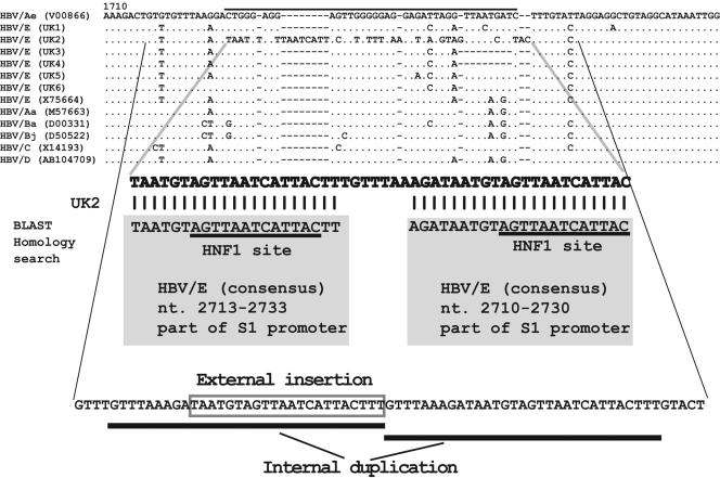
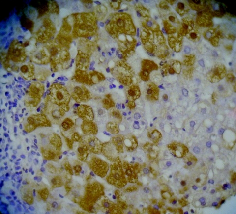
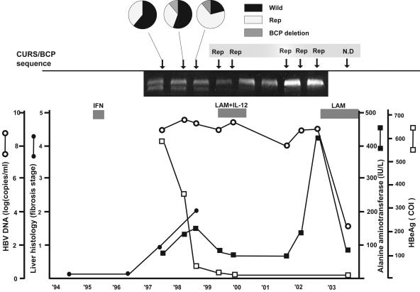
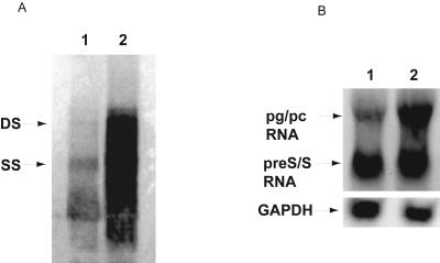
The genetic diversity of hepatitis B virus (HBV) strains has evolved through mutations such as point mutations, deletions or insertions, and recombination. We identified and characterized a novel type of mutation which is a complex of external insertion, deletion, and internal duplication in sequences from one of six patients with chronic hepatitis B virus genotype E (HBV/E). We provisionally named this mutation a "replacement mutation"; the core promoter upstream regulatory sequence/basic core promoter was replaced with a part of the S1 promoter covering the hepatocyte nuclear factor 1 (HNF1) binding site, followed by a tandem repeat of the HNF1 site. A longitudinal analysis of the HBV population over 6 years showed the clonal change from wild-type HBV/E to replacement-mutant type, resulting in a lower hepatitis B (HB) e antigen titer, a high HBV DNA level in serum, and progression of liver fibrosis. In an in vitro study using a replication model, the replacement-mutant HBV showed higher replication levels than the wild-type HBV/E replicon, probably mediated by altered transcription factor binding. Additionally, this HNF1 site replacement mutation was associated with excessive HB nucleocapsid protein expression in hepatocytes, in both in vivo and in vitro studies. This novel mutation may be specific to HBV genotype E, and its prevalence requires further investigation.


Figures

FIG. 1.
FIG. 1.
Nucleotide alignment of the six HBV genotype E sequences of the core upstream regulatory region to the basic core promoter region, with references to other genotypes and subtypes. The sequence enlarged under the alignment shows the UK2-specific 50-nucleotide sequences which were analyzed by BLAST homology search. The 1st to 21st nucleotides and 29th to 50th nucleotides matched the conserved sequences of HBV genotype E, nt 2713 to 2733 and nt 2710 to 2730, shown in shaded rectangles. Furthermore, the consequences of mutations which occurred in the UK2 sequence are shown. The normal CURS/BCP sequences are deleted, the S1 promoter, including the HNF1 site, is externally inserted, and internal duplication occurs.
FIG. 2.
FIG. 2.
Immunohistochemical detection of HBcAg in the liver of the patient 2. Strong staining is observed in both nuclei and cytoplasm.
FIG. 3.
FIG. 3.
Clinical course of patient 2 based on HBV DNA levels, fibrosis stage, HBeAg titer, and ALT levels. The three circular charts show how the proportions of wild type, replacement mutation (Rep) type, and BCP deletion type changed during the time course. The electropherogram (5%) shows that mixed types (wild type and Rep) are found during the first three points. ○, HBV DNA levels; •, fibrosis stage; □, HBeAg titer; ▪, ALT levels. The patient was treated with IFN monotherapy and then with the combination therapy of lamivudine (LAM) and interleukin-12 (IL-12). The patient has been treated with LAM since 2003. COI, cutoff index.
FIG. 4.
FIG. 4.
(A) Southern blot analysis of intracellular replication competence of replacement mutation with HNF1 site. Double-stranded (DS) DNA and single-stranded (SS) DNA are indicated by arrows. The pUC19-HBV/E replacement replicon (lane 2) replicates at a much higher rate than the pUC19-HBV/E wild-type replicon (lane 1). (B) Northern blot analysis of HBV transcripts. Lane 1, pUC19-HBV/E wild-type replicon; lane 2, pUC19-HBV/E replacement replicon. pg/pc RNA, pregenomic/precore RNA. A GAPDH probe was used to quantify RNA in each lane.
FIG. 5.
FIG. 5.
Intracellular localization of HBcAg (magnification, ×400). Immunofluorescence staining of HBcAg in HuH-7 cells transfected with (a) pUC19-HBV/E wild-type replicon and (c) pUC19-HBV/E replacement replicon. (b and d) Nuclear staining for the same set of cells was performed by use of DNA staining. DAPI stain shows as red.

References

    1. Abe, A., K. Inoue, T. Tanaka, J. Kato, N. Kajiyama, R. Kawaguchi, S. Tanaka, M. Yoshiba, and M. Kohara. 1999. Quantitation of hepatitis B virus genomic DNA by real-time detection PCR. J. Clin. Microbiol. 37:2899-2903. - PMC - PubMed
    1. Ahn, S. H., A. Kramvis, S. Kawai, H. C. Spangenberg, J. Li, G. Kimbi, M. Kew, J. Wands, and S. Tong. 2003. Sequence variation upstream of precore translation initiation codon reduces hepatitis B virus e antigen production. Gastroenterology 125:1370-1378. - PubMed
    1. Altschul, S. F., T. L. Madden, A. A. Schaffer, J. Zhang, Z. Zhang, W. Miller, and D. J. Lipman. 1997. Gapped BLAST and PSI-BLAST: a new generation of protein database search programs. Nucleic Acids Res. 25:3389-3402. - PMC - PubMed
    1. Bhat, R. A., P. P. Ulrich, and G. N. Vyas. 1990. Molecular characterization of a new variant of hepatitis B virus in a persistently infected homosexual man. Hepatology 11:271-276. - PubMed
    1. Blum, H. E., Z.-S. Zhang, E. Galun, F. von Weizsäcker, B. Garner, T. J. Liang, and J. R. Wands. 1992. Hepatitis B virus X protein is not central to the viral life cycle in vitro. J. Virol. 66:1223-1227. - PMC - PubMed

Publication types

LinkOut - more resources