

Histology-based expression profiling yields novel prognostic markers in human glioblastoma
- PMID: 16254489
- PMCID: PMC1557632
- DOI: 10.1097/01.jnen.0000186940.14779.90
Histology-based expression profiling yields novel prognostic markers in human glioblastoma
Abstract
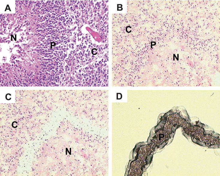
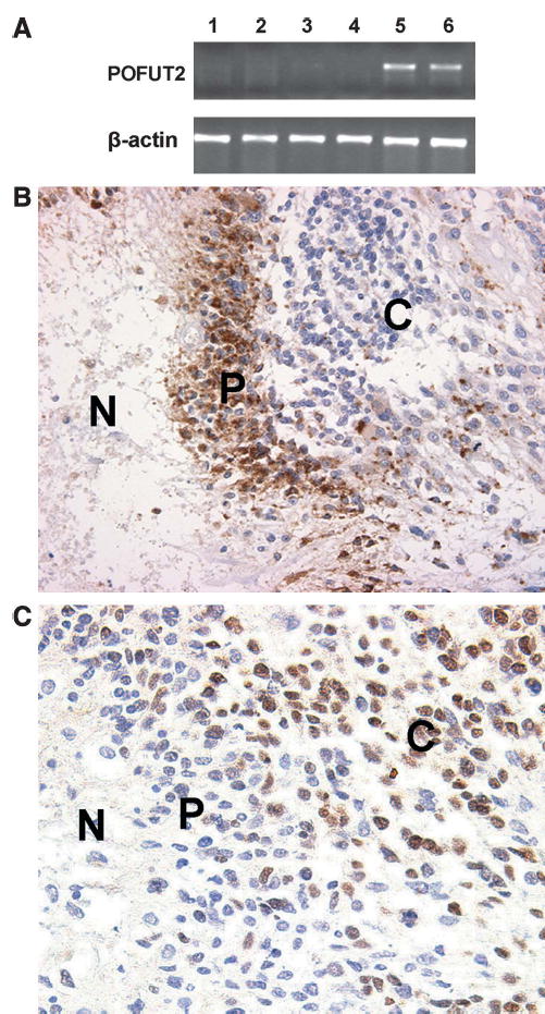
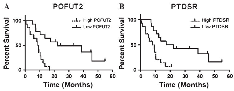
Although the prognosis for patients with glioblastoma is poor, survival is variable, with some patients surviving longer than others. For this reason, there has been longstanding interest in the identification of prognostic markers for glioblastoma. We hypothesized that specific histologic features known to correlate with malignancy most likely express molecules that are directly related to the aggressive behavior of these tumors. We further hypothesized that such molecules could be used as biomarkers to predict behavior in a manner that might add prognostic power to sole histologic observation of the feature. We reasoned that perinecrotic tumor cell palisading, which denotes the most aggressive forms of malignant gliomas, would be a striking histologic feature on which to test this hypothesis. We therefore used laser capture microdissection and oligonucleotide arrays to detect molecules differentially expressed in perinecrotic palisades. A set of RNAs (including POFUT2, PTDSR, PLOD2, ATF5, and HK2) that were differentially expressed in 3 initially studied, microdissected glioblastomas also provided prognostic information in an independent set of 28 glioblastomas that did not all have perinecrotic palisades. On validation in a second, larger independent series, this approach could be applied to other human glioma types to derive tissue biomarkers that could offer ancillary prognostic and predictive information alongside standard histopathologic examination.
Figures
References
-
- Jaffe ES, Harris NL, Stein H, et al. World Health Organization Classification of Tumours of Haematopoietic and Lymphoid Tissues Lyon: WHO/IARC, 2001
-
- Fletcher CDM, Unni KK, Mertens F. World Health Organization Classification of Tumours of Soft Tissue and Bone Lyon: WHO/IARC, 2002
-
- Nutt CL, Mani DR, Betensky RA, et al. Gene expression-based classification of malignant gliomas correlates better with survival than histological classification. Cancer Res. 2003;63:1602–7. - PubMed
-
- Sasaki H, Zlatescu MC, Betensky RA, et al. Histopathological–molecular genetic correlations in referral pathologist-diagnosed low-grade ‘oligo-dendroglioma. ’ J Neuropathol Exp Neurol. 2002;61:58–63. - PubMed
Publication types
MeSH terms
Substances
Grants and funding
LinkOut - more resources
Full Text Sources
Other Literature Sources
Medical
Molecular Biology Databases
Miscellaneous
