

The interphotoreceptor retinoid-binding protein (IRBP) of the chicken (Gallus gallus domesticus)
- PMID: 16254552
- PMCID: PMC2895308
The interphotoreceptor retinoid-binding protein (IRBP) of the chicken (Gallus gallus domesticus)
Abstract
Purpose: Despite decades of investigation, the function of interphotoreceptor retinoid binding protein (IRBP), the most abundant protein in the interphotoreceptor matrix of vertebrates, remains enigmatic. Roles for IRBP in the visual cycle of rod photoreceptors and in the independent visual cycle of cone photoreceptors have been suggested, yet very little is known of the biology of IRBP in cone-dominant retinas, such as those of diurnal birds. Our aim was to identify and characterize expression of the IRBP of the cone-dominant chicken (Gallus gallus domesticus).
Methods: Chicken IRBP mRNA was identified by PCR cloning. Primary protein structure, genomic organization, and phylogenies were determined through comparative sequence analyses. Expression of IRBP mRNA was characterized by northern analysis and by in situ hybridization on cryosectioned chicken retina. Expression of the IRBP protein was characterized by western blotting and by indirect immunofluorescence on cryosectioned retina and on retinal whole mounts.
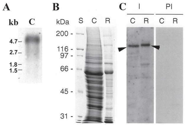
Results: The chicken IRBP gene encodes a secreted protein with a predicted 1,252 amino acid length. The gene structure for chicken IRBP resembles that of most other vertebrates, with four homologous, modular repeats and introns within only the fourth module. Each module is more homologous with the corresponding module in other species than it is with the remaining chicken modules. Chicken retinal tissue contains a single IRBP mRNA transcript of approximately 4.8 kb and western analysis of chicken retina shows a single major band of 140 kDa. Chicken IRBP mRNA is expressed exclusively by retinal photoreceptor cells and the intensity of the hybridization signal shows light/dark rhythmicity. The IRBP protein is localized to the interphotoreceptor matrix of the chicken retina and to intracellular regions of photoreceptors, with a spatial distribution indicating an association with cone outer segments.
Conclusions: The high degree of conservation of IRBP's primary structure, genomic organization, and cell-specific expression within the retinas of all vertebrates examined to date, including those with cone-dominant retinas, implies a conserved role for IRBP in photoreceptor function and/or health. Expression of chicken IRBP and its mRNA are functionally regulated. This report provides a necessary first step to explore a specific function for IRBP in the cone visual cycle.
Figures


Similar articles
-
Cone outer segment extracellular matrix as binding domain for interphotoreceptor retinoid-binding protein.J Comp Neurol. 2012 Mar 1;520(4):756-69. doi: 10.1002/cne.22773. J Comp Neurol. 2012. PMID: 21935947 Free PMC article.
-
Cone outer segment and Müller microvilli pericellular matrices provide binding domains for interphotoreceptor retinoid-binding protein (IRBP).Exp Eye Res. 2013 Aug;113:192-202. doi: 10.1016/j.exer.2013.02.003. Epub 2013 Mar 5. Exp Eye Res. 2013. PMID: 23470504
-
Goldfish cones secrete a two-repeat interphotoreceptor retinoid-binding protein.J Mol Evol. 1995 Nov;41(5):646-56. doi: 10.1007/BF00175823. J Mol Evol. 1995. PMID: 7490779
-
Interphotoreceptor retinoid-binding protein (IRBP). Molecular biology and physiological role in the visual cycle of rhodopsin.Mol Neurobiol. 1993 Spring;7(1):61-85. doi: 10.1007/BF02780609. Mol Neurobiol. 1993. PMID: 8318167 Review.
-
Interphotoreceptor retinoid-binding protein: biochemistry and molecular biology.Prog Clin Biol Res. 1991;362:115-37. Prog Clin Biol Res. 1991. PMID: 2003123 Review.
Cited by
-
Cone outer segment extracellular matrix as binding domain for interphotoreceptor retinoid-binding protein.J Comp Neurol. 2012 Mar 1;520(4):756-69. doi: 10.1002/cne.22773. J Comp Neurol. 2012. PMID: 21935947 Free PMC article.
-
Circadian regulation of vertebrate cone photoreceptor function.Elife. 2021 Sep 22;10:e68903. doi: 10.7554/eLife.68903. Elife. 2021. PMID: 34550876 Free PMC article.
-
Module structure of interphotoreceptor retinoid-binding protein (IRBP) may provide bases for its complex role in the visual cycle - structure/function study of Xenopus IRBP.BMC Biochem. 2007 Aug 4;8:15. doi: 10.1186/1471-2091-8-15. BMC Biochem. 2007. PMID: 17683573 Free PMC article.
-
Evolution and expression of the phosphodiesterase 6 genes unveils vertebrate novelty to control photosensitivity.BMC Evol Biol. 2016 Jun 13;16(1):124. doi: 10.1186/s12862-016-0695-z. BMC Evol Biol. 2016. PMID: 27296292 Free PMC article.
-
Interphotoreceptor retinoid-binding protein gene structure in tetrapods and teleost fish.Mol Vis. 2006 Dec 9;12:1565-85. Mol Vis. 2006. PMID: 17200656 Free PMC article.
References
-
- Fernald RD. Evolution of eyes. Curr Opin Neurobiol. 2000;10:444–50. - PubMed
-
- Hageman GS, Johnson LV. Structure, composition and function of the retinal interphotoreceptor matrix. In: Osborne NN, Chader GJ, editors. Progress in retinal research. Vol. 10. Oxford: Pergamon Press; 1990. pp. 207–49.
-
- Mieziewska K. The interphotoreceptor matrix, a space in sight. Microsc Res Tech. 1996;35:463–71. - PubMed
-
- Hageman GS, Kuehn MH. Biology of the interphotoreceptor matrix-retinal interface. In: Marmor MF, Wolfensberber TJ, editors. The retinal pigment epithelium: function and disease. New York: Oxford University Press; 1998. pp. 417–54.
-
- Hollyfield JG. Hyaluronan and the functional organization of the interphotoreceptor matrix. Invest Ophthalmol Vis Sci. 1999;40:2767–9. - PubMed
Publication types
MeSH terms
Substances
Grants and funding
LinkOut - more resources
Full Text Sources
