. 2005 Dec 12;493(2):177-92.
doi: 10.1002/cne.20711.

Postnatal phenotype and localization of spinal cord V1 derived interneurons

Affiliations

Postnatal phenotype and localization of spinal cord V1 derived interneurons

Francisco J Alvarez et al. J Comp Neurol. .

Abstract

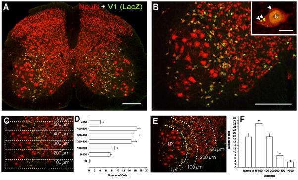
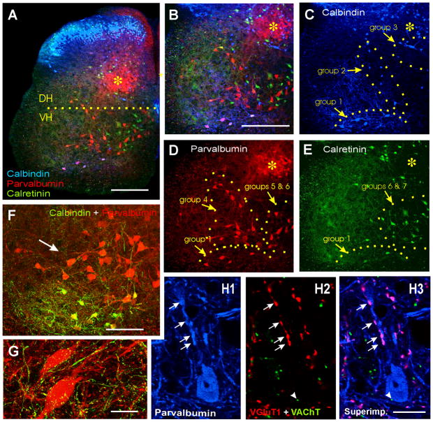
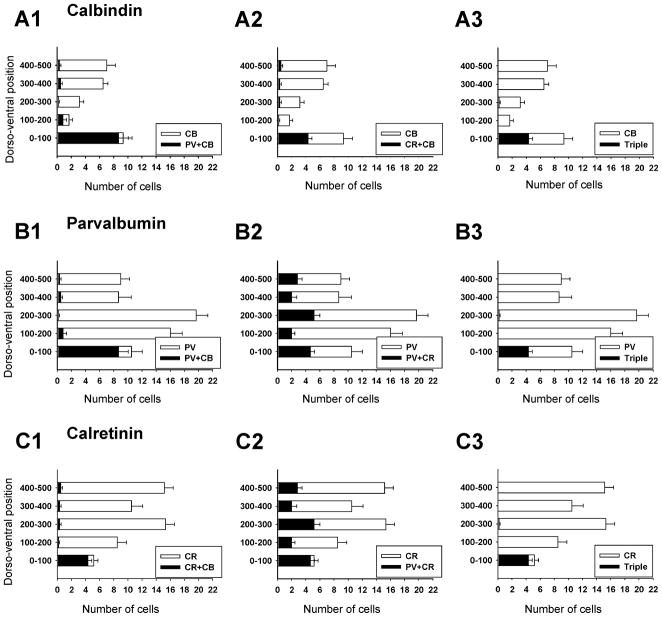
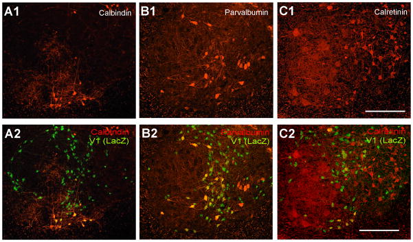
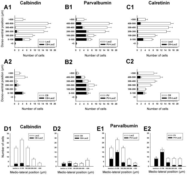
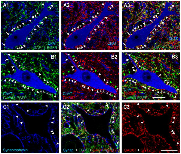
Developmental studies identified four classes (V0, V1, V2, V3) of embryonic interneurons in the ventral spinal cord. Very little is known, however, about their adult phenotypes. Therefore, we characterized the location, neurotransmitter phenotype, calcium-buffering protein expression, and axon distributions of V1-derived neurons in the adult mouse spinal cord. In the mature (P20 and older) spinal cord, most V1-derived neurons are located in lateral LVII and in LIX, few in medial LVII, and none in LVIII. Approximately 40% express calbindin and/or parvalbumin, while few express calretinin. Of seven groups of ventral interneurons identified according to calcium-buffering protein expression, two groups (1 and 4) correspond with V1-derived neurons. Group 1 are Renshaw cells and intensely express calbindin and coexpress parvalbumin and calretinin. They represent 9% of the V1 population. Group 4 express only parvalbumin and represent 27% of V1-derived neurons. V1-derived Group 4 neurons receive contacts from primary sensory afferents and are therefore proprioceptive interneurons. The most ventral neurons in this group receive convergent calbindin-IR Renshaw cell inputs. This subgroup resembles Ia inhibitory interneurons (IaINs) and represents 13% of V1-derived neurons. Adult V1-interneuron axons target LIX and LVII and some enter the deep dorsal horn. V1 axons do not cross the midline. V1-derived axonal varicosities were mostly (>80%) glycinergic and a third were GABAergic. None were glutamatergic or cholinergic. In summary, V1 interneurons develop into ipsilaterally projecting, inhibitory interneurons that include Renshaw cells, Ia inhibitory interneurons, and other unidentified proprioceptive interneurons.


Figures

Figure 1
Figure 1. Distribution of V1-derived interneurons in the spinal cord
A, Shows a low magnification view of a section through a P20 V1-lacZ mouse spinal cord immunolabeled for βgal (FITC, green) and NeuN (Cy3, red). V1 lacZ expressing neurons are yellow. βgal-IR V1-derived interneurons are restricted to the ventral horn. B, Shows the distribution of V1-derived interneurons at higher magnification. Most V1-interneurons are concentrated in an arch in LVII medial to LIX motoneuron pools (larger neurons in the image). Inset at top right corner shows a high magnification of a NeuN-IR V1-derived interneuron. βgal-IR is concentrated in the nucleus (N) and in intracytoplasmic inclusions of variable sizes (arrowheads). C, D, Dorso-ventral distribution of V1-derived interneurons. C is the same image as in B but with overlays indicating dorso-ventral bins where neurons were counted. Only neurons with a clear immunolabeled nucleus (both NeuN and β-gal-IR) and containing a nucleolus were counted. D shows the dorso-ventral spread of V1-derived interneurons. Y-axis = 0 represents the border between the ventral horn and the ventral funiculus, bins are 100 μm of increasing distance from this border (see C). Although V1-derived interneurons are dispersed throughout the whole dorso-ventral extent of the ventral horn they are more abundant in the dorsal half. Data represent the average number of neurons in each bin per ventral horn in 40 μm thick sections (n = 8 ventral horns from two different animals, error bars represent SEM). E, F, Medio-lateral distribution of V1-derived interneurons analyzed in the same sections. In F the X-axis represents medio-lateral distribution. 0 represents the border between LVII and lateral LIX. Each bin represents 100 μm incremental distances from this border (see E). Most V1-interneurons were located in LIX or within 200 μm of LIX motoneuron pools. Very few neurons were located medially. Scale Bars: A,B, 200 μm; inset, 10 μm.
Figure 2
Figure 2. Calbindin, parvalbumin and calretinin immunoreactivities in ventral interneurons
A, Low magnification image of a triple immunolabeled section: calbindin-IR (blue, Cy5), parvalbumin-IR (red, Cy3) and calretinin (green, FITC). The horizontal line extends from the dorsal tip of the central canal line and indicates the approximate boundary between ventral (VH) and dorsal (DH) horns. The region labeled with an asterisk indicates a region in medial LV and LVI containing a dense plexus of parvalbumin-IR muscle afferent fibers and many parvalbumin-IR neurons. This region is also labeled for orientation purposes in following images at higher magnification. B, Triple immunolabeled ventral horn. C, D, E, Show each immunoreaction separately. Panels B and C–E are at the same magnification. Immunolabeled ventral interneurons were divided into 7 groups according to localization and expression of the different calcium buffering proteins (see text and table 2). F, Higher magnification image of parvalbumin-IR only neurons (group 4) and below Renshaw cells that coexpress parvalbumin and calbindin (yellow). Calbindin-IR axons (i.e. Renshaw cell projections) surround many parvalbunin-IR neurons. G, High magnification 2D projection of the full 3D reconstruction (12 confocal planes separated by 1.5 μm) of two parvalbumin-IR neurons (red, Cy3) located in group 4 and surrounded by calbindin-IR varicose fibers (green, FITC). This image is representative of the high density of calbindin-IR contacts found around these neurons. H1–3, A parvalbumin-IR neuron in group 4 displaying contacts on its dendrites and soma from parvalbumin-IR boutons (arrows in H1) that are also VGluT1-IR (arrows in H2; colocalization in pink, H3). These presumably belong to muscle primary sensory afferents. In addition, this cell is sparsely contacted by cholinergic (VAChT-IR) varicosities (arrowhead). H1 shows parvalbumin-IR only (blue, Cy5); H2, VGluT1 (red, Cy3) and VAChT (green, FITC); H3 all three superimposed. Scale Bars: A,B, 200 μm; F, 100 μm; G,H, 20 μm.
Figure 3
Figure 3. Dorso-ventral distribution of calbindin, parvalbumin and calretinin ventral interneurons
A, Distribution of neurons that contained only calbindin-IR (white bars) or co-localized calbindin and parvalbumin (black bars in A1) or calbindin and calretinin (black bars in A2) or all three (black bars in A3). Most calbindin-IR neurons are located in the ventral-most 100 mm of the ventral horn (group 1). Significant co-localization is found in this cell group: 92% also contained parvalbumin and 46% calretinin. This group of cells is the only one in the ventral horn showing significant co-expression of all three calcium buffering proteins (e.g. A3, B3, C3). Few calbindin-IR neurons are found in the mid ventral horn and their density increases again close to the dorsal horn (upper bins). Virtually none of these more dorsally located calbindin-IR neurons co-expressed parvalbumin or calretinin. B, Distribution of neurons that expressed only parvalbumin-IR (white bars), or co-localized calbindin (A1), calretinin (A2) or both (A3) with parvalbumin. In contrast to calbindin-IR neurons, most parvalbumin-IR neurons are located in the mid ventral horn regions (distances 100 to 300 μm from the ventral funiculus). There is only limited co-localization with calbindin (19.6% of all parvalbumin-IR neurons in the ventral horn) except for Renshaw cells (first 100 μm bin). Similarly, 44% of parvalbumin-IR neurons co-expressed calretinin in the Renshaw cell bin, while in more dorsal bins the percentage varied from 12 to 31%. Most neurons with co-localized parvalbumin and calretinin immunoreactivities were located quite medially (group 6). C, Distribution of neurons with only calretinin-IR (white bars) and co-localization of calretinin with calbindin (A1), parvalbumin (A2) or both (A3). Calretinin-IR neurons are distributed throughout the whole dorso-ventral extent of the ventral horn, but most are quite medially located. Apart from the Renshaw cell group very little co-localization was found with calbindin and co-localization with parvalbumin varied from 18% to 33% of neurons in bins above the Renshaw cell group. The numerical data were obtained from triple-immunolabeled sections taken from 6 ventral horns at the L5 level. One example is shown in Figure 2. Y-axis indicates dorso-ventral location; 0 is the border with the ventral funiculus. X-axis is the average number of cells per ventral horn counted. Error bars indicate SEM.
Figure 4
Figure 4. Expression of calbindin, parvalbumin or calretinin in V1-derived interneurons
A, Ventral horn dual-immunolabeled for calbindin and βgal shown with calbindin-immunoreactivity only (A1) and βgal superimposed (A2). B, C, Similar images for parvalbumin and βgal (B1,2) and calretinin and βgal (C1,2). All ventral calbindin-IR neurons (group 1; Renshaw cells) were βgal-IR. Similarly, most parvalbumin-IR neurons close to LIX (groups 1 and 4) were also βgal-IR. Dorsomedial calbindin-IR neurons (group 3), most calretinin neurons (groups 6 and 7) and ventromedial parvalbumin neurons (groups 5 and 6) were almost never βgal-IR. Around half of calbindin-IR neurons located in mid-regions of the ventral horn (group 2) were βgal-IR. Quantitative data from these preparations are shown in Figure 5. Scale Bars in C1 and C2, 200 μm.
Figure 5
Figure 5. Distributions of V1-derived interneurons expressing calcium-buffering proteins
A,B,C, Dorso-ventral distributions of βgal-IR neurons expressing calbindin (A), parvalbumin (B) or calretinin (C). Most V1-derived neurons expressing calbindin and calretinin are located in the first 100 μm bin from the ventral funiculus, while parvalbumin-IR neurons are distributed throughout the dorso-ventral extension of the ventral horn. Many V1-derived interneurons, particularly those located in more dorsal bins, did not express any calcium buffering protein. D,E, Medio-lateral distribution of βgal-IR neurons expressing calbindin (D) or parvalbumin (E). V1-neurons containing calretinin largely coincide with group 1 calbindin/parvalbumin neurons and therefore were not analyzed further. Medio-lateral distribution histograms show that most (75% to 90% in different bins) calbindin and/or parvalbumin expressing neurons in LIX or LVII close to LIX, are V1-derived. In contrast, medially located calbindin-IR or parvalbumin-IR neurons were rarely V1-derived (3% to 14% in at distances greater than 200 μm from the LIX border). Data collected from 5 and 7 ventral horns sampled from βgal/calbindin and βgal/parvalbumin immunolabeled sections respectively. Error bars indicate SEMs.
Figure 6
Figure 6. Ia inhibitory interneurons are derived from V1-interneurons
A, Low magnification image showing parvalbumin-IR neurons in blue (Cy5), calbindin-IR neurons in red (Cy3) and βgal-immunoreactivity in green (FITC). Almost all ventrally located neurons in red (calbindin only) or pink (calbindin + parvalbumin co-expression) are V1-derived (arrowheads) and correspond to Renshaw cells (group 1). Dorsally located calbindin-IR neurons (red) do not co-express parvalbumin and are not V1-derived. Blue neurons (parvalbumin-only) in close spatial relationship to LIX (group 4) are V1 derived, while more medially located parvalbumin-IR neuron are not. A dense plexus of calbindin-IR fibers extends from the Renshaw cell area into LIX and also into the LVII region that contains many V1-derived parvalbumin-IR neurons (arrows). B, High magnification of mid LVII showing V1-derived (βgal-IR) parvalbumin-IR neurons (blue) receiving a dense innervation from calbindin-IR fibers (red). C, A similar image to B, but from ventral LVII and including one Renshaw cell co-expressing both calbindin and parvalbumin (pink). Calbindin-IR contacts were more numerous on V1-derived parvalbumin-only neurons than on Renshaw cells or other neurons that lacked parvalbumin or calbindin (immunostained in other preparations with NeuN, not shown). Almost all neurons densely innervated by calbindin-IR fibers were parvalbumin-IR and V1-derived. These neurons likely correspond with Ia inhibitory interneurons (see text and Figure 2). B and C are superimpositions of 3 confocal planes separated by 1 μm. Scale bars: A, 100 μm; B,C, 10 μm.
Figure 7
Figure 7. V1 axon projections in the spinal cord
A,B, Low magnification images of adult thoracic spinal cord (>3 months old) immunostained for NeuN (Cy3, red, shown alone in A) and EGFP containing V1 axons (GAP43-EGFP, FITC, green, B). A, laminar boundaries and the locations of the dorsal columns (DC), the lateral funiculus (LF), dorsolateral funiculus (DLF) and ventral funiculus (VF) are indicated based on cytoarchitectonic criteria. Clarke’s column, the origin of the dorsal spinocerebellar tract (DCST), is also outlined (cc). B, shows the distribution of EGFP V1 axons. V1 axons are abundant in the ventral spinal cord (LVII–LIX) and in lateral LV–LVI, but not in more dorsal laminae or in Clarke’s column (medial LV–LVI). In the white matter V1 axons are very abundant in the lateral and ventral funiculus in proximity to the ventral horn (blue dotted line is at the gray/white matter boundary) and in a prominent dorsolateral funiculus bundle. There are no fibers in the dorsal columns and V1 axons are sparse in more peripheral regions of the lateral and ventral funiculi. C, High magnification image of large NeuN labeled DSCT neurons and smaller interneurons in LVII (one of them contains weak EGFP immunoreactivity in the cytoplasm) surrounded by V1-derived EGFP-IR axons. The density of EGFP fibers and varicosities drops dramatically in Clarke’s column (CC) and there are very few direct contacts on the cell bodies. The varicosities labeled as c1 and c2 are shown at higher magnification in the insets. They appear separated by small gaps from the somatic membrane of the large DSCT neurons. See for comparison direct appositions on LVII interneurons shown in c3. In conclusion, direct V1 contacts onto DSCT neurson occur at densities lower than on LVII interneurons or LIX motoneurons (see panel F). This image is a superimposition of two confocal images separated by 0.5 μm in the z-axis. D, High magnification image of ventral midline region (note central canal dorsally) showing the ventral white commissure (VWC) virtually free of labeled axons and none that appear crossing. The mid-ventral edge of the spinal cord is indicated by a dotted blue line. E, Medium magnification of the adult (>3 months old) spinal cord in lumbar 4, immunostained for NeuN (Cy3, red) and EGFP V1 axons (FITC, green). The distribution of V1 axons is similar at all spinal cord levels (see A,B). LVIII is prominent in this segment and shows less V1 axons than other adjacent ventral horn laminae. Asterisk indicates the medial LV–LVI region in continuity with Clarke’s column of thoracic segments. This region receives abundant innervation from Ia, Ib and II fibers (see Figure 2A) and has a low density of V1 axons. F, High magnification confocal image of large NeuN-immunoreactive neurons in LIX (motoneurons) densely innervated by EGFP-immunoreactive varicosities of V1 origin. The area labeled with an arrowhead is shown at high magnification in the inset. V1 EGFP-IR varicosities make direct contacts onto the cell bodies of motoneurons. Scale bars: A,E, 200 μm; C, F, 20 μm, insets in C and F 5 μm; D, 50 μm.
Figure 8
Figure 8. V1 axons express markers indicating a glycinergic and/or GABA phenotype
A, High magnification single confocal optical section through a motoneuron cell body (ChAT-IR, blue, Cy5) surrounded by V1-derived axons (GAP43-EGFP, green, Alexa 488 in A1) and GlyT2-IR axons (Cy3, red, A2). Both images are superimposed in E3. White arrowheads indicate V1-varicosities that also contain GlyT2-immunoreactivity and are in contact with the motoneuron. Grey arrowheads indicate some GlyT2-IR varicosities in contact with the motoneuron that are not V1-derived (GAP43-EGFP negative). Arrows point to ChAT-IR C-terminals, none of which were V1-derived. Most V1-derived contacts on motoneurons express GlyT2-IR suggesting a glycinergic phenotype. B, High magnification single confocal optical section through another motoneuron in a section stained for GAD67-immunoreactivity. A proportion of V1 derived varicosities were GAD67-IR (white arrowheads) but there were also significant numbers of GAD67-IR terminals that were not V1-derived (grey arrowheads) and many V1-derived varicosities that did not contain GAD67-IR (green arrowheads). ChAT-IR C-terminals (one marked with an arrow) were not V1-derived or GAD67-IR. C, A region of lamina IX neuropil with unstained motoeneuron cell bodies and proximal dendrites and triple immunolabeled for synaptophysin (blue, Cy5), EGFP V1 axons (Alexa 488, green) and GAD67-GlyT2, both antibodies detected with the same fluorochrome (TRITC, red). C1 shows synaptophysin immunolabeling alone. C2, EGFP and GAD67/GlyT2 immunostaining superimposed. C3, GAD67/GlyT2 immunoreactivities. Note that some boutons are filled with immunoreactivity due to intense GAD67 and others are only labeled in the periphery, as characteristic for GlyT2. Arrowheads are in the same positions in C1, C2, and C3 and indicate V1 EGFP positive varicosities containing synaptophysin (i.e., synaptic sites). All V1 synaptic sites contain some red immunofluorescence indicating that they are GAD67 and/or GlyT2 immunoreactive and therefore inhibitory. Similar analyses were performed in random regions of LIX and LVII neuropil with similar results (not shown in figure). Scale bars, B3, C3, 20 μm. Panels A nad B are at the same magnification.

References

    1. Altman J, Bayer SA. Development of the Human Spinal Cord. Oxford University Press Inc; New York: 2001.
    1. Alvarez FJ, Dewey DE, Harrington DA, Fyffe RE. Cell-type specific organization of glycine receptor clusters in the mammalian spinal cord. J Comp Neurol. 1997;379:150–170. - PubMed
    1. Alvarez FJ, Dewey DE, McMillin P, Fyffe RE. Distribution of cholinergic contacts on Renshaw cells in the rat spinal cord: a light microscopic study. J Physiol (Lond) 1999;15:787–797. - PMC - PubMed
    1. Alvarez FJ, Jonas P, Sapir T, Geiman EJ, Hartley R, Todd AJ, Goulding M. Abstract Viewer/Itinerary Planner. Washington, DC: Society for Neuroscience; 2003. Postnatal phenotype and localization of spinal cord V1 derived interneurons. Program No. 39.3. Online.
    1. Alvarez FJ, Villalba RM, Zerda R, Schneider SP. Vesicular glutamate transporters in the spinal cord, with special reference to sensory primary afferent synapses. J Comp Neurol. 2004;472:257–280. - PubMed

Publication types