. 2005 Nov 1;77(21):6902-6908.
doi: 10.1021/ac050963m.

Analysis of biogenic amine variability among individual fly heads with micellar electrokinetic capillary chromatography-electrochemical detection

Affiliations

Analysis of biogenic amine variability among individual fly heads with micellar electrokinetic capillary chromatography-electrochemical detection

Paula R Powell et al. Anal Chem. .

Abstract

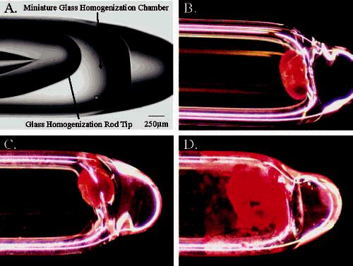
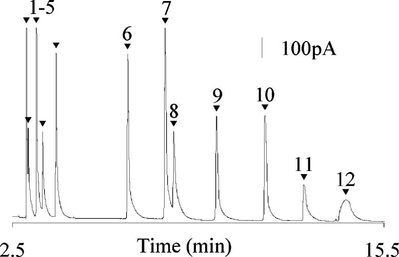
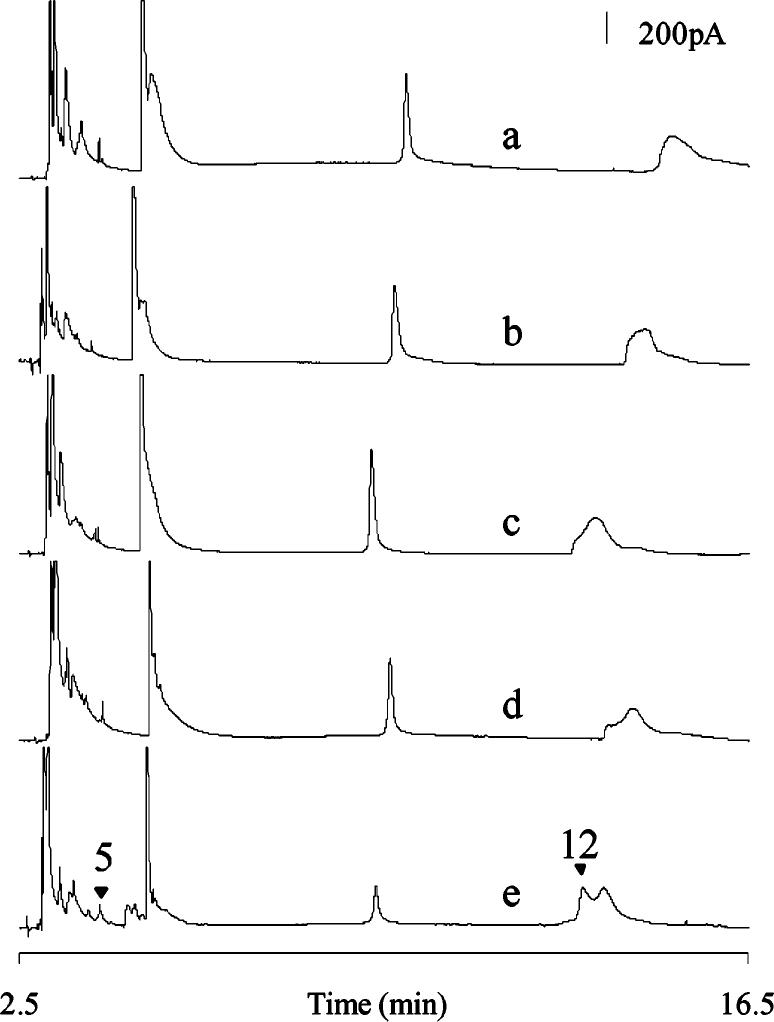
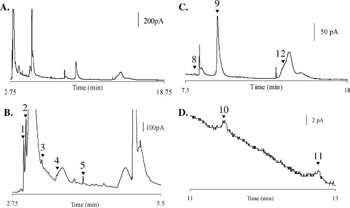
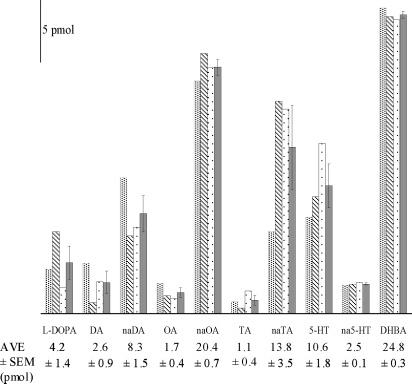
Neurochemical variability among individual Drosophila heads has been examined with the sensitivity of electrochemical detection and the selectivity of micellar electrokinetic capillary chromatography. Homogenization of single Drosophila heads in volumes as small as 100 nL has been accomplished. Here we demonstrate reproducible separations for single fly heads in 250-nL volumes providing a 4-fold increase in sensitivity without overloading the electrochemical detector. This increase in sensitivity allows detection of previously undetected analytes, such as N-acetyltyramine (naTA) and octopamine (OA). Analytes including L-3,4-dihydroxyphenylalanine, N-acetyl octopamine, N-acetyldopamine, naTA, N-acetylserotonin, OA, dopamine, tyramine, and serotonin also have been consistently identified in single-head homogenates and observed with homogenates representing populations of Drosophila. Neurochemical variation between individual flies as well as the consistency within a population indicates varying amounts of neurotransmitter turnover. The inception, design, and fabrication of a miniature tissue homogenizer has enabled the separation of biogenic amines and metabolites from these severely volume-limited single Drosophila head homogenates.


Figures

Figure 1
Figure 1
(A) Image of miniature tissue homogenizer with arrows indicating a glass pipet pulled and sealed serving as the homogenization chamber and a glass capillary pulled and sealed serving as a tissue homogenizing rod. (B) Glass microvial containing a single fly head. (C) Glass microvial containing a single fly head in 250 nL of homogenization solution before homogenization. (D) Glass microvial containing a single fly head in 250 nL of homogenization solution after homogenization. Scale bar is the same for all four panels.
Figure 2
Figure 2
Separation of 100 μM concentration of each amine: l-DOPA (1), naOA (2), naDA (3), naTA (4), na5-HT (5), NE (6), E (7), OA (8), DHBA (9), DA (10), TA (11), and 5-HT (12). Separation was performed in a 13-μm-i.d. capillary with a separation potential of 562 V/cm. The working electrode was held at +750 mV vs a Ag/AgCl reference electrode. Buffer: 10 mM TES, 30 mM SDS, and 2% 1-propanol.
Figure 3
Figure 3
Electropherograms of single Drosophila head homogenates in (a) 1000, (b) 750, (c) 500, (d) 250, and (e) 100 nL of homogenization buffer. Highlighted are na5-HT (5) and 5-HT (12) corresponding to the standards labeled in Figure 2. Separations were performed on unique 13-μm-i.d. capillaries with separation potentials of (a) 566, (b) 568, (c) 558, (d) 552, and (e) 563 V/cm. Unique working electrodes were used for each separation and were held at +750 mV vs a Ag/AgCl reference electrode. Buffer: 10 mM TES, 30 mM SDS, and 2% 1-propanol.
Figure 4
Figure 4
Electropherogram of a single fly head homogenate in 250 nL of homogenization solution. (A) MEKC–EC separation of a single male head homogenate. (B) Enlargement of the first 5 min of the separation highlighting l-DOPA (1), naOA (2), naDA (3), naTA (4), and na5-HT (5). (C) Enlargement of the latter half of the separation highlighting OA (8), DHBA (9), and 5-HT (12). (D) Enlargement emphasizing DA (10) and TA (11). Field strength for the 13-μm-i.d. capillary is 568 V/cm. Working electrode was held at +750 mV vs a Ag/AgCl reference electrode. Buffer: 10 mM TES, 30 mM SDS, and 2% 1-propanol.
Figure 5
Figure 5
Quantification of neurochemicals from three single fly heads at 250-nL volumes injected from a micrometer-sized tissue homogenizer. Bars represent values for individual flies 1, 2, and 3 (patterns) and the average (solid) ± SEM of the amines in the three flies. Numeric averages and SEM are reported below each amine. Separations were performed in a 13-mm-i.d. capillary with separation potentials of (1) 568, (2) 568, and (3) 552 V/cm. The working electrodes were held at +750 mV vs a Ag/AgCl reference electrode. These numbers have been calculated based on the known concentration of the corresponding analyte in the standard mixture separated prior to the homogenate and the values normalized to the internal standard DHBA.

References

    1. Brown AS, Gershon S. J. Neural Transm. Gen. Sect. 1993;91:75–109. - PubMed
    1. Miyamoto S, Duncan GE, Marx CE, Lieberman JA. Mol. Psychiatry. 2005;10:79–104. - PubMed
    1. Ogawa N. Neurology. 1998;51:S13–20. - PubMed
    1. Stoof JC, Winogrodzka A, van Muiswinkel FL, Wolters EC, Voorn P, Groenewegen HJ, Booij J, Drukarch B. Eur. J. Pharmacol. 1999;375:75–86. - PubMed
    1. Powell PR, Ewing AG. Anal. Bioanal. Chem. 2005;382:581–591. - PubMed

Publication types

MeSH terms

Substances

LinkOut - more resources