

A detailed characterization of loud noise stress: Intensity analysis of hypothalamo-pituitary-adrenocortical axis and brain activation
- PMID: 16256084
- PMCID: PMC2409188
- DOI: 10.1016/j.brainres.2005.09.031
A detailed characterization of loud noise stress: Intensity analysis of hypothalamo-pituitary-adrenocortical axis and brain activation
Abstract
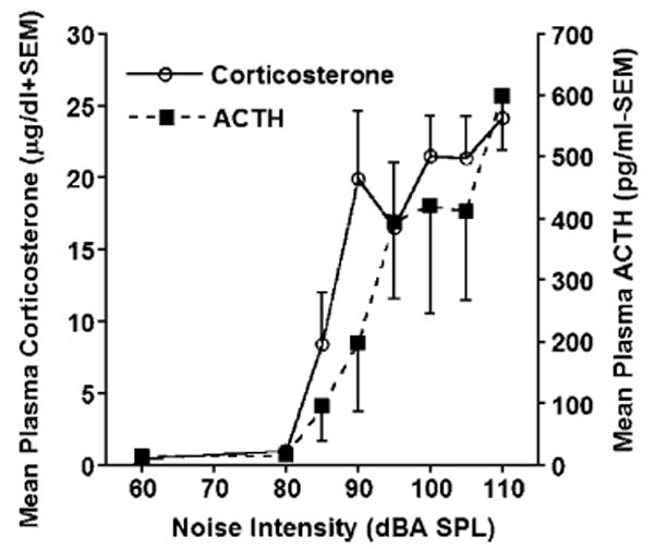
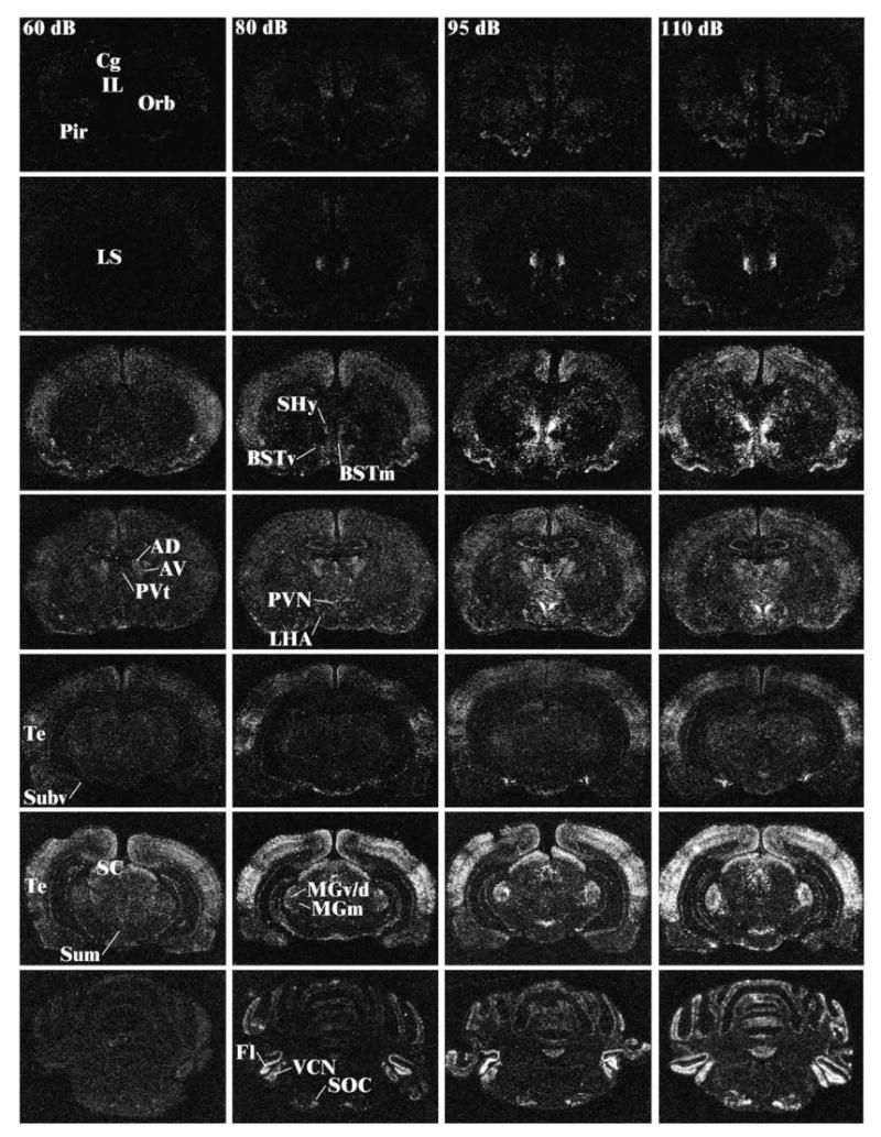
The present studies were undertaken to help determine the putative neural circuits mediating activation of the hypothalamo-pituitary-adrenocortical (HPA) axis and the release of adrenocorticotropin hormone (ACTH) and corticosterone in response to the perceived threat of loud noise. This experiment involved placing rats in acoustic chambers overnight to avoid any handling and context changes prior to noise exposure, which was done for 30 min (between 9:00 and 10:00 am) at intensities of 80, 85, 90, 95, 100, 105, and 110 dBA in different groups (n = 8), and included a background condition (60 dBA ambient noise). This manipulation produced a noise-intensity-related increase in plasma ACTH and corticosterone levels, with levels beginning to rise at approximately 85 dBA. c-fos mRNA induction was very low in the brains of the control and 80 dBA groups, but several brain regions displayed a noise-intensity-related induction. Of these, several forebrain regions displayed c-fos mRNA induction highly correlated (r > 0.70) with that observed in the paraventricular hypothalamic nucleus and plasma ACTH levels. These regions included the ventrolateral septum, the anteroventral subiculum, several preoptic nuclei, the anterior bed nucleus of the stria terminalis (BNST), the anterior paraventricular nucleus of the thalamus, and the medial subdivision of the medial geniculate body. Together with prior findings with audiogenic stress, the present results suggest that either or both the anterior BNST or the lateral septum is ideally situated to trigger HPA axis activation by stimuli that are potentially threatening.
Figures
Similar articles
-
Cannabinoid receptor type 1 antagonism significantly modulates basal and loud noise induced neural and hypothalamic-pituitary-adrenal axis responses in male Sprague-Dawley rats.Neuroscience. 2012 Mar 1;204:64-73. doi: 10.1016/j.neuroscience.2011.11.043. Epub 2011 Nov 28. Neuroscience. 2012. PMID: 22138156 Free PMC article.
-
Lesions of the medial geniculate nuclei specifically block corticosterone release and induction of c-fos mRNA in the forebrain associated with audiogenic stress in rats.J Neurosci. 1997 Aug 1;17(15):5979-92. doi: 10.1523/JNEUROSCI.17-15-05979.1997. J Neurosci. 1997. PMID: 9221794 Free PMC article.
-
Remote CB1 receptor antagonist administration reveals multiple sites of tonic and phasic endocannabinoid neuroendocrine regulation.Psychoneuroendocrinology. 2020 Mar;113:104549. doi: 10.1016/j.psyneuen.2019.104549. Epub 2019 Dec 19. Psychoneuroendocrinology. 2020. PMID: 31884322 Free PMC article.
-
The anteroventral bed nucleus of the stria terminalis differentially regulates hypothalamic-pituitary-adrenocortical axis responses to acute and chronic stress.Endocrinology. 2008 Feb;149(2):818-26. doi: 10.1210/en.2007-0883. Epub 2007 Nov 26. Endocrinology. 2008. PMID: 18039788 Free PMC article.
-
The cochlea as an independent neuroendocrine organ: expression and possible roles of a local hypothalamic-pituitary-adrenal axis-equivalent signaling system.Hear Res. 2012 Jun;288(1-2):3-18. doi: 10.1016/j.heares.2012.03.007. Epub 2012 Mar 29. Hear Res. 2012. PMID: 22484018 Free PMC article. Review.
Cited by
-
Prenatal stress dysregulates resting-state functional connectivity and sensory motifs.Neurobiol Stress. 2021 May 27;15:100345. doi: 10.1016/j.ynstr.2021.100345. eCollection 2021 Nov. Neurobiol Stress. 2021. PMID: 34124321 Free PMC article.
-
Vacuum-cleaner noise and acute stress responses in female C57BL/6 mice (Mus musculus).J Am Assoc Lab Anim Sci. 2010 May;49(3):300-6. J Am Assoc Lab Anim Sci. 2010. PMID: 20587160 Free PMC article.
-
Gum chewing inhibits the sensory processing and the propagation of stress-related information in a brain network.PLoS One. 2013;8(4):e57111. doi: 10.1371/journal.pone.0057111. Epub 2013 Apr 3. PLoS One. 2013. PMID: 23573184 Free PMC article.
-
Cannabinoid receptor type 1 antagonism significantly modulates basal and loud noise induced neural and hypothalamic-pituitary-adrenal axis responses in male Sprague-Dawley rats.Neuroscience. 2012 Mar 1;204:64-73. doi: 10.1016/j.neuroscience.2011.11.043. Epub 2011 Nov 28. Neuroscience. 2012. PMID: 22138156 Free PMC article.
-
Chronic stress from adolescence to adulthood increases adiposity and anxiety in rats with decreased expression of Krtcap3.Front Genet. 2024 Jan 23;14:1247232. doi: 10.3389/fgene.2023.1247232. eCollection 2023. Front Genet. 2024. PMID: 38323241 Free PMC article.
References
-
- Abercrombie ED, Keefe KA, DiFrischia DS, Zigmond MJ. Differential effect of stress on in vivo dopamine release in striatum, nucleus accumbens, and medial frontal cortex. J Neurochem. 1989;52:1655–1658. - PubMed
-
- Antoni FA. Hypothalamic control of adrenocorticotropin secretion: advances since the discovery of 41-residue corticotropin-releasing factor. Endocr Rev. 1986;7:351–378. - PubMed
-
- Brake WG, Flores G, Francis D, Meaney MJ, Srivastava LK, Gratton A. Enhanced nucleus accumbens dopamine and plasma corticosterone stress responses in adult rats with neonatal excitotoxic lesions to the medial prefrontal cortex. Neuroscience. 2000;96:687–695. - PubMed
-
- Briese E, Cabanac M. Stress hyperthermia: physiological arguments that it is a fever. Physiol Behav. 1991;49:1153–1157. - PubMed
-
- Buller KM, Day TA. Systemic administration of interleukin-1beta activates select populations of central amygdala afferents. J Comp Neurol. 2002;452:288–296. - PubMed
Publication types
MeSH terms
Substances
Grants and funding
LinkOut - more resources
Full Text Sources
Medical
