. 2005 Oct 24;2005(5):249-55.
doi: 10.1155/MI.2005.249.

Inhibitors of mitogen-activated protein kinases downregulate COX-2 expression in human chondrocytes

Affiliations

Inhibitors of mitogen-activated protein kinases downregulate COX-2 expression in human chondrocytes

Riina Nieminen et al. Mediators Inflamm. .

Abstract

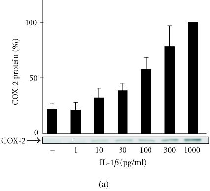
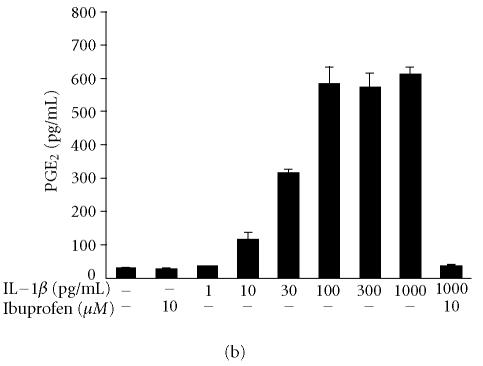
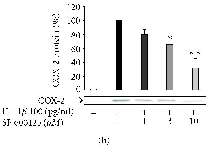
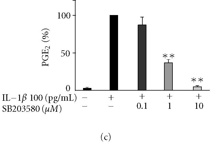
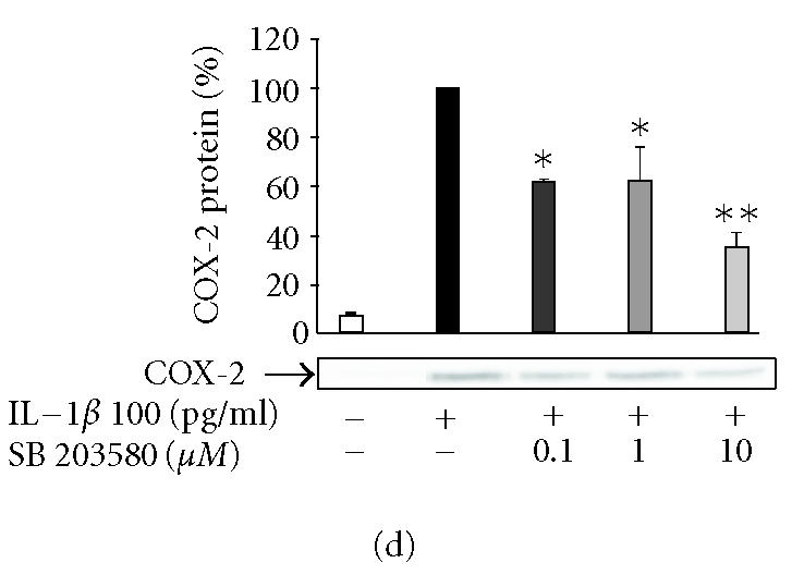
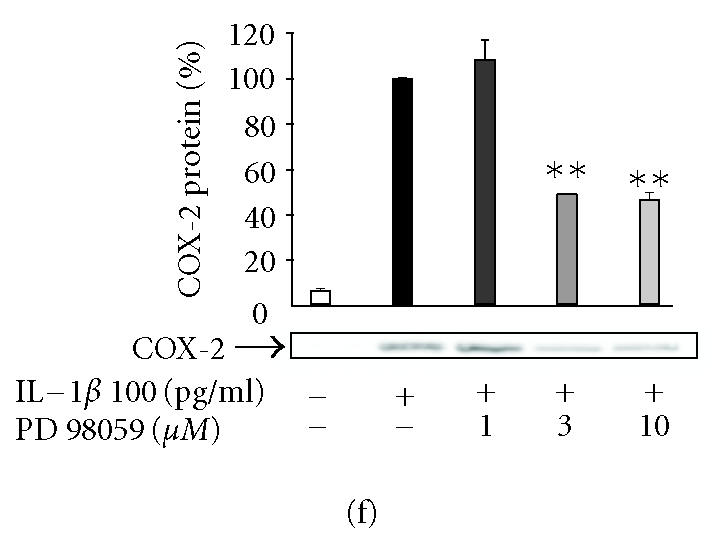
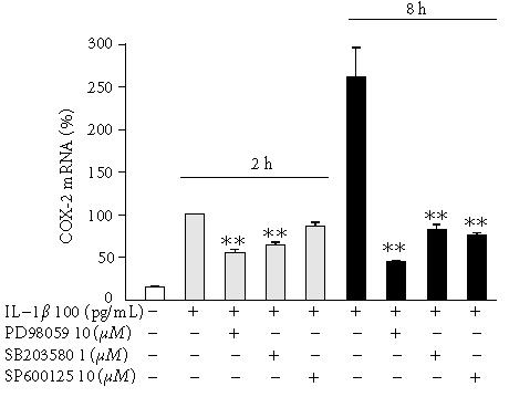
Inducible prostaglandin synthase (cyclooxygenase-2, COX-2) is expressed in rheumatoid and osteoarthritic cartilage and produces high amounts of proinflammatory prostanoids in the joint. In the present study we investigated the effects of the inhibitors of mitogen-activated protein kinase (MAPK) pathways Erk1/2, p38, and JNK on COX-2 expression and prostaglandin E2 (PGE2) production in human chondrocytes. Proinflammatory cytokine IL-1beta caused a transient activation of Erk1/2, p38, and JNK in immortalized human T/C28a2 chondrocytes and that was followed by enhanced COX-2 expression and PGE2 production. PD98059 (an inhibitor of Erk1/2 pathway) suppressed IL-1-induced COX-2 expression and PGE2 production in a dose-dependent manner, and seemed to have an inhibitory effect on COX-2 activity. SB203580 (an inhibitor of p38 pathway) but not its negative control compound SB202474 inhibited COX-2 protein and mRNA expression and subsequent PGE2 synthesis at micromolar drug concentrations. SP600125 (a recently developed JNK inhibitor) but not its negative control compound N1-methyl-1,9-pyrazolanthrone downregulated COX-2 expression and PGE2 formation in a dose-dependent manner. SP600125 did not downregulate IL-1-induced COX-2 mRNA expression when measured 2 h after addition of IL-1beta but suppressed mRNA levels in the later time points suggesting post-transcriptional regulation. Our results suggest that activation of Erk1/2, p38, and JNK pathways belongs to the signaling cascades that mediate the upregulation of COX-2 expression and PGE2 production in human chondrocytes exposed to proinflammatory cytokine IL-1beta.


Figures

Figure 1
Figure 1
The effects of IL-1β on JNK, p38, and Erk1/2 MAPK activation in human T/C28a2 chondrocytes. The chondrocytes were stimulated with IL-1β (100 pg/mL). Incubations were terminated at the indicated time points. Two parallel immunoblots were run from same cell lysates using antibodies against the Thr-183/Tyr-185, Thr-180/Tyr-182, and Thr-202/Tyr-204 phosphorylated (ie, activated) JNK (p-JNK), p38 (p-p38), and Erk1/2 (p-Erk1/2) and against total JNK, p38, and Erk1/2. The experiment was repeated three times with similar results.
Figure 2
Figure 2
The effects of IL-1β on COX-2 protein expression and PGE2 production in human T/C28a2 chondrocytes. (a) Human chondrocytes were incubated for 24 h in the presence of increasing concentrations of IL-1β, and COX-2 protein was measured by Western blot. (b) Human chondrocytes were incubated for 24 h in the presence of increasing concentrations of IL-1β, and PGE2 concentrations in the culture medium were measured by radioimmunoassay. COX inhibitor ibuprofen (10 μM) was used as a control compound. Mean ± SEM, n = 4 − 6. In (a), a representative gel is shown under the bars.
Figure 2
Figure 2
The effects of IL-1β on COX-2 protein expression and PGE2 production in human T/C28a2 chondrocytes. (a) Human chondrocytes were incubated for 24 h in the presence of increasing concentrations of IL-1β, and COX-2 protein was measured by Western blot. (b) Human chondrocytes were incubated for 24 h in the presence of increasing concentrations of IL-1β, and PGE2 concentrations in the culture medium were measured by radioimmunoassay. COX inhibitor ibuprofen (10 μM) was used as a control compound. Mean ± SEM, n = 4 − 6. In (a), a representative gel is shown under the bars.
Figure 3
Figure 3
The effects of SP600125, SB203580, and PD98059 on PGE2 production and COX-2 protein expression in IL-1β-stimulated human T/C28a2 chondrocytes. Human chondrocytes were incubated with IL-1β (100 pg/mL) and increasing concentrations of (a), (b) SP600125 (JNK inhibitor), (c), (d) SB203580 (p38 inhibitor), and (e), (f) PD98059 (Erk1/2 inhibitor). After 24 h, incubations were terminated, and PGE2 concentrations in the culture medium were measured by RIA ((a), (c), (e)) and COX-2 protein was measured by Western blot ((b), (d), (f)). (g) SP600125 (10 μM), SB203580 (1 μM), and PD98059 (10 μM) were added to the cell culture at the same time (0 h) or 6 h after IL-1β (6 h). After 24 h, PGE2 concentrations were measured in the culture medium by RIA. Mean ± SEM, n = 4, ∗∗ indicates P < .01 as compared with the respective control. In (b), (d), and (f) a representative gel is shown under the bars.
Figure 3
Figure 3
The effects of SP600125, SB203580, and PD98059 on PGE2 production and COX-2 protein expression in IL-1β-stimulated human T/C28a2 chondrocytes. Human chondrocytes were incubated with IL-1β (100 pg/mL) and increasing concentrations of (a), (b) SP600125 (JNK inhibitor), (c), (d) SB203580 (p38 inhibitor), and (e), (f) PD98059 (Erk1/2 inhibitor). After 24 h, incubations were terminated, and PGE2 concentrations in the culture medium were measured by RIA ((a), (c), (e)) and COX-2 protein was measured by Western blot ((b), (d), (f)). (g) SP600125 (10 μM), SB203580 (1 μM), and PD98059 (10 μM) were added to the cell culture at the same time (0 h) or 6 h after IL-1β (6 h). After 24 h, PGE2 concentrations were measured in the culture medium by RIA. Mean ± SEM, n = 4, ∗∗ indicates P < .01 as compared with the respective control. In (b), (d), and (f) a representative gel is shown under the bars.
Figure 4
Figure 4
The effects of SP600125, SB203580, and PD98059 on COX-2 mRNA levels in IL-1β-stimulated human chondrocytes. Human chondrocytes were incubated with IL-1β (100 pg/ml) and with or without SP600125, SB203580, and PD98059. Incubations were terminated at the indicated time points, and the extracted total RNA was subjected to real-time RT-PCR. COX-2 mRNA levels were normalized against β-actin mRNA. Mean ± SEM, n = 6, ∗∗ indicates P < .01 as compared with cells treated with IL-1β only.

Similar articles

Cited by

References

    1. Dubois RN, Abramson SB, Crofford L, et al. Cyclooxygenase in biology and disease. FASEB J. 1998;12(12):1063–1073. - PubMed
    1. Needleman P, Turk J, Jakschik BA, Morrison AR, Lefkowith JB. Arachidonic acid metabolism. Annu Rev Biochem. 1986;55:69–102. - PubMed
    1. Turini ME, DuBois RN. Cyclooxygenase-2: a therapeutic target. Annu Rev Med. 2002;53:35–57. - PubMed
    1. Vane JR, Bakhle YS, Botting RM. Cyclooxygenases 1 and 2. Annu Rev Pharmacol Toxicol. 1998;38:97–120. - PubMed
    1. Amin AR, Attur M, Patel RN, et al. Superinduction of cyclooxygenase-2 activity in human osteoarthritis-affected cartilage. Influence of nitric oxide. J Clin Invest. 1997;99(6):1231–1237. - PMC - PubMed

MeSH terms