. 2005 Oct 31;33(19):6277-86.
doi: 10.1093/nar/gki947. Print 2005.

The POU protein Oct-6 is a nucleocytoplasmic shuttling protein

Affiliations

The POU protein Oct-6 is a nucleocytoplasmic shuttling protein

Constanze Baranek et al. Nucleic Acids Res. .

Abstract

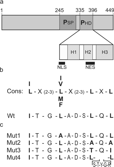
Like many POU domain proteins, Oct-6 plays important roles during vertebrate development. In accord with its function as a transcriptional regulator during neurogenesis and myelination, Oct-6 is predominantly found in the nucleus. Nuclear import is mediated by a nuclear localization signal at the N-terminal end of the POU homeodomain. Here we show, that Oct-6 in addition contains a nuclear export signal so that Oct-6 is able to shuttle constantly between nucleus and cytoplasm. This nuclear export signal is also localized in the POU homeodomain as part of helix 2 and the connecting loop to DNA recognition helix 3. It conforms to the consensus of hydrophobic leucine-rich export sequences and mediates export from the nucleus via CRM1/Exp1. Several amino acid substitutions or insertions that inactivate this nuclear export sequence, reduce DNA-binding of Oct-6 to its octamer recognition element slighty, but interfere strongly with Oct-6-dependent transcriptional activation, thus arguing that nuclear export and nucleocytoplasmic shuttling are essential aspects of Oct-6 function. Importantly, the nuclear export signal identified for Oct-6 is conserved in most, if not all other vertebrate POU proteins. Nuclear export might therefore be of general relevance for POU protein function throughout development.


Figures

Figure 1
Figure 1
Identification and mutation of the putative NES of Oct-6. (a) schematic representation of Oct-6 with its POU domain consisting of POU-specific domain (PSP) and POU homeodomain (PHD) with its 3 helices H1, H2 and H3. Locations of the previously identified NLS (14) and the putative NES within the POU homeodomain are highlighted. (b) comparison of the putative NES in Oct-6 (Wt, amino acids 367–376) with the consensus sequence (cons) of leucine-rich NES (16). (c) Mutations introduced into the NES of Oct-6 and used throughout this study.
Figure 2
Figure 2
Characterization of GFP–Oct-6 and GFP-Oct-6-POU. (a) schematic representation of the GFP fusion constructs. (b) western blot analyses of extracts from COS-7 cells expressing either Oct-6 (Wt), GFP–Oct-6 or GFP-Oct-6-POU (GFP-POU) using anti-Oct-6 (α-Oct-6) or anti-GFP (α-GFP) antibodies. Protein sizes are indicated on the right side of the panels. (−), control COS-7 extract. (c) Subcellular localization of GFP–Oct-6 in transiently transfected NIH 3T3 cells by direct fluorescence microscopy.
Figure 3
Figure 3
Oct-6 shuttles between the nucleus and the cytoplasm as revealed by heterokaryon assays. NIH 3T3 cells were transfected with expression plasmids for GFP-SOX10 (a) GFP-Sox10-ΔNES (b) GFP–Oct-6 (c) and GFP-Oct-6-POU (d) before they were subjected to heterokaryon formation with HeLa cells. After 2 h, cells were fixed, counterstained with DAPI (middle panel) and analyzed for GFP autofluorescence (left panel). Right panels show the heterokaryons in phase contrast. Donor mouse nuclei are marked by an arrowhead and recipient human nuclei by asterisks.
Figure 4
Figure 4
The leucine-rich sequence in the POU homeodomain of Oct-6 is required for nucleocytoplasmic shuttling. NIH 3T3 cells were transfected with expression plasmids for wild-type (Wt) GFP-Oct-6-POU (a), and versions which carried the Mut1 (b), the Mut2 (c), the Mut3 (d) or the Mut4 (e) mutation in their POU domain (see also Figure 1c). After cycloheximide treatment, transfected NIH 3T3 cells were subjected to heterokaryon formation with HeLa cells. After 2 h, cells were fixed, counterstained with DAPI (middle panel) and analyzed for GFP autofluorescence (left panel). Right panels show the heterokaryons in phase contrast. Donor mouse nuclei are marked by an arrowhead and recipient human nuclei by asterisks.
Figure 5
Figure 5
The hydrophobic leucine-rich sequence in Oct-6 functions as a NES when transferred to a heterologous protein. Purified GST–GFP chimeric proteins were microinjected into the nuclei of HeLa cells together with rabbit Cy3-coupled IgG as a marker for the nuclear injection site. 45 min after injection, the cells were fixed and counterstained with DAPI (last row). The injected rabbit IgG was detected exclusively within the nucleus (second row), whereas the GST–GFP chimeras containing either the NES of Sox10 (b, Sox10-NES) or of Oct-6 (c, Oct-6 NES) were significantly translocated from the nuclear injection site into the cytoplasm as revealed by the green autofluorescence of the GFP moiety (first row). In contrast those GST–GFP chimeras containing mutant versions of the Sox10 NES (a, Sox10-ΔNES) or the Oct-6 NES (d, Mut1-NES; e, Mut2-NES) failed to leave the microinjected nucleus. Merged pictures of the upper two rows are shown in the third row.
Figure 6
Figure 6
The NES in the POU domain is the dominant NES of Oct-6. NIH 3T3 cells were transfected with expression plasmids for wild-type (Wt) GFP–Oct-6 (a) and versions which carried the Mut1 (b) or the Mut2 (c) mutation. After cycloheximide treatment, transfected NIH 3T3 cells were subjected to heterokaryon formation with HeLa cells. After 2 h, cells were fixed, counterstained with DAPI (middle panel) and analyzed for GFP autofluorescence (left panel). Right panels show the heterokaryons in phase contrast. Donor mouse nuclei are marked by an arrowhead and recipient human nuclei by asterisks.
Figure 7
Figure 7
Nucleocytoplasmic shuttling of Oct-6 is CRM1/Exp1-dependent. (a) NIH 3T3 cells were transfected with an expression plasmid for GFP–Oct-6 and pretreated with leptomycin B, before they were subjected to heterokaryon formation with HeLa cells. After 2 h in the continued presence of leptomycin B, cells were fixed, counterstained with DAPI (middle panel) and analyzed for GFP autofluorescence (left panel). Right panels show the heterokaryons in phase contrast. (b) A GST–GFP chimera carrying the NES of Oct-6 was microinjected into the nuclei of leptomycin B-pretreated HeLa cells together with rabbit Cy3-coupled IgG as a marker for the nuclear injection site. Cells were kept under leptomycin B. 45 min after injection, the cells were fixed and counterstained with DAPI (middle row). The injected rabbit IgG (right panel) as well as the GST–GFP chimera carrying the NES of Oct-6 (left panel) were detected exclusively within the nucleus.
Figure 8
Figure 8
The NES modulates the transcriptional activity of Oct-6. (a) western blot analysis of COS-7 extracts containing wild-type (Wt) GFP-Oct-6-POU or its mutant versions Mut1, Mut2, Mut3 and Mut4 using an anti-Oct-6 antibody. Approximately equal amounts of each Oct-6 protein were loaded. (b) EMSA of wild-type (Wt) GFP-Oct-6-POU and its mutant versions Mut1, Mut2, Mut3 and Mut4 using HSVoct as probe. In both (a) and (b), the relative amount of each protein is indicated below the lanes. The highest amount of wild-type protein used in the electrophoretic mobility shift assay was arbitrarily set to 1. (c–e) Transient transfections in U138 and Neuro2a cells with a luciferase reporter under the control of the HSVoct-binding site and the β-globin minimal promoter (HSVoct-luc). The luciferase reporter was transfected either alone or in combination with Oct-6 in its wild-type form (Wt) or in mutant versions (Mut1, Mut2, Mut3 and Mut4). Whereas 0.5 µg of Oct-6 expression plasmids were used in (c), increasing amounts (20 ng, 0.2 µg, 0.5 µg, 1 µg and 2.5 µg) were used in (d). Cells in (e) were kept in the absence (− LMB) or presence (+ LMB) of leptomycin B after transfection. Data from at least two independent experiments each performed in duplicates are presented as fold inductions ± SEM with the activity for each luciferase reporter in the absence of co-transfected Oct-6 arbitrarily set to 1.
Figure 9
Figure 9
Conservation of the Oct-6 export sequence in other homeodomain proteins. (a) Comparison of the NES defined in this work with the corresponding regions of POU proteins representative of classes I–VI and select other homeodomain proteins. Bcd, Bicoid; En-2, Engrailed-2. (b) Nucleocytoplasmic shuttling activity of the Brn-4 POU domain. NIH 3T3 cells were transfected with an expression plasmid for wild-type GFP-Brn-4-POU. After cycloheximide treatment, transfected NIH 3T3 cells were subjected to heterokaryon formation with HeLa cells. After 2 h, cells were fixed, counterstained with DAPI (middle panel) and analyzed for GFP autofluorescence (left panel). Right panels show the heterokaryons in phase contrast. Donor mouse nuclei are marked by an arrowhead and recipient human nuclei by an asterisk.

References

    1. Ryan A.K., Rosenfeld M.G. POU domain family values: flexibility, partnerships, and developmental codes. Genes Dev. 1997;11:1207–1225. - PubMed
    1. Wegner M., Drolet D.W., Rosenfeld M.G. POU-domain proteins: structure and function of developmental regulators. Curr. Opin. Cell Biol. 1993;5:488–498. - PubMed
    1. Hara Y., Rovescalli A.C., Kim Y., Nirenberg M. Structure and evolution of four POU domain genes expressed in mouse brain. Proc. Natl Acad. Sci. USA. 1992;89:3280–3284. - PMC - PubMed
    1. Alvarez-Bolado G., Rosenfeld M.G., Swanson L.W. Model of forebrain regionalization based on spatiotemporal patterns of POU-III homeobox gene expression, birthdates, and morphological features. J. Comp. Neurol. 1995;355:237–295. - PubMed
    1. Bermingham J.R., Jr, Scherer S.S., O'Connell S., Arroyo E., Kalla K.A., Powell F.L., Rosenfeld M.G. Tst-1/Oct-6/SCIP regulates a unique step in peripheral myelination and is required for normal respiration. Genes Dev. 1996;10:1751–1762. - PubMed

Publication types