. 2005 Nov 7;202(9):1171-7.
doi: 10.1084/jem.20050630. Epub 2005 Oct 31.

RNA-associated autoantigens activate B cells by combined B cell antigen receptor/Toll-like receptor 7 engagement

Affiliations

RNA-associated autoantigens activate B cells by combined B cell antigen receptor/Toll-like receptor 7 engagement

Christina M Lau et al. J Exp Med. .

Abstract

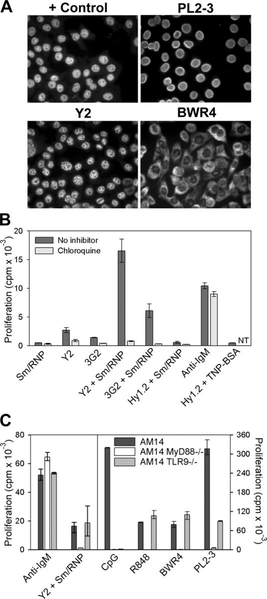
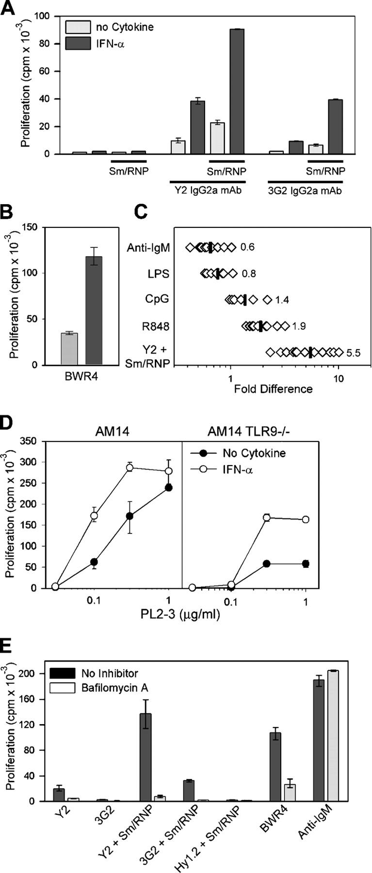
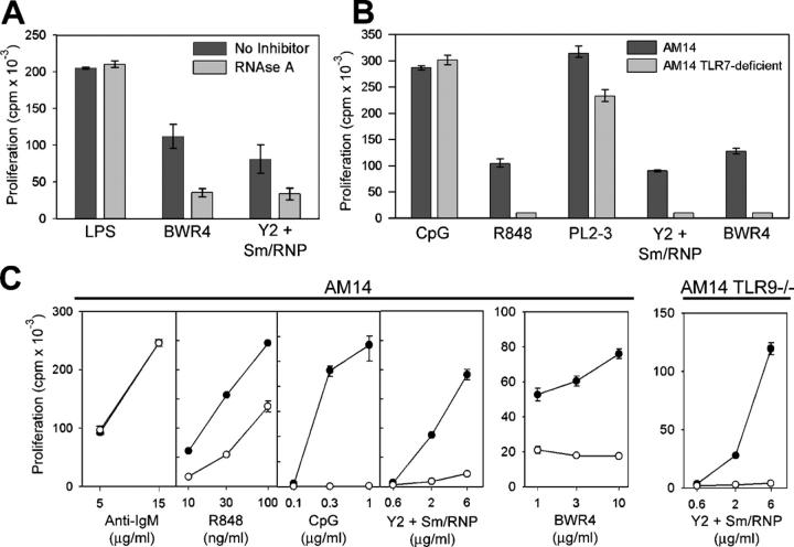
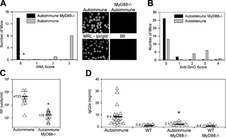
Previous studies (Leadbetter, E.A., I.R. Rifkin, A.H. Hohlbaum, B. Beaudette, M.J. Shlomchik, and A. Marshak-Rothstein. 2002. Nature. 416:603-607; Viglianti, G.A., C.M. Lau, T.M. Hanley, B.A. Miko, M.J. Shlomchik, and A. Marshak-Rothstein. 2003. Immunity. 19:837-847) established the unique capacity of DNA and DNA-associated autoantigens to activate autoreactive B cells via sequential engagement of the B cell antigen receptor (BCR) and Toll-like receptor (TLR) 9. We demonstrate that this two-receptor paradigm can be extended to the BCR/TLR7 activation of autoreactive B cells by RNA and RNA-associated autoantigens. These data implicate TLR recognition of endogenous ligands in the response to both DNA- and RNA-associated autoantigens. Importantly, the response to RNA-associated autoantigens was markedly enhanced by IFN-alpha, a cytokine strongly linked to disease progression in patients with systemic lupus erythematosus (SLE). As further evidence that TLRs play a key role in autoantibody responses in SLE, we found that autoimmune-prone mice, lacking the TLR adaptor protein MyD88, had markedly reduced chromatin, Sm, and rheumatoid factor autoantibody titers.


Figures

Figure 1.
Figure 1.
RNA-associated ICs activate AM14 B cells by a TLR-dependent mechanism. (A) HEp2 cells were stained with MRL/lpr serum (control) or the mAbs PL2-3, Y2, or BWR4. (B) AM14 B cells were stimulated with the anti-Sm mAbs Y2 and 3G2 or the anti-TNP mAb Hy1.2, alone or in combination with Sm/RNP antigen, with 5 μg/ml anti-IgM F(ab′)2 or with Hy1.2 + TNP BSA ICs in the presence or absence of chloroquine. Data shown are representative of between three and four experiments. NT, not tested. (C) AM14, AM14 MyD88-deficient, and AM14 TLR9-deficient B cells were stimulated with 5 μg/ml anti-IgM F(ab′)2, Sm/RNP IC, 1 μg/ml CpG s-ODN 1826, 10 ng/ml R848, BWR4, or PL2-3. Data shown are representative of at least two experiments per group. Values in B and C indicate means ± SD.
Figure 2.
Figure 2.
Sm/RNP IC and BWR4 activation of AM14 B cells is enhanced by IFN-α. Proliferative response of AM14 B cells to anti-Sm mAbs or Sm/RNP ICs (A) or BWR4 (B) in the presence or absence of 1,000 U/ml of IFN-α. Values indicate means ± SD. (C) Ratio of the response to anti-IgM F(ab′)2, LPS, CpG, R848, or Sm/RNP ICs stimulated in the presence or absence of IFN-α. Each point represents data from one experiment; data includes AM14 and AM14 TLR9-deficient responder populations. Mean values are indicated by vertical lines. (D) Response of AM14 and AM14 TLR9-deficient B cells to PL2-3 in the presence or absence of 1000 U/ml of IFN-α. (E) Stimulation of AM14 B cells by the indicated ligands after preincubation with IFN-α in the presence or absence of bafilomycin A. Values in D and E indicate means ± SD.
Figure 3.
Figure 3.
Sm/RNP IC and BWR4 activation of AM14 B cells depends on RNA activation of TLR7 and is blocked by s-ODN 2088. (A) AM14 B cells were stimulated with Y2 + Sm/RNP, BWR4, or LPS in the presence or absence of RNase A. Shown are the means of three experiment per group ± SD. (B) AM14 and AM14 TLR7-deficient B cells were stimulated with 1 μg/ml CpG, 10 ng/ml R848, PL2-3, or Y2 + Sm/RNP or BWR4. Data shown are representative of two experiments. (C) AM14 and AM14 TLR9-deficient B cells were stimulated with anti-IgM F(ab′)2, R848, CpG, Y2 + Sm/RNP, or BWR4 in the presence (○) or absence (•) of 4 μg/ml s-ODN 2088 and are representative of between four and seven experiments per group. In all cases, the Y2 + Sm/RNP– and BWR4-stimulated cells were preincubated with IFN-α. Values in B and C indicate means ± SD.
Figure 4.
Figure 4.
Absence of autoantibody production in MyD88-deficient mice. (A) ANA production by 20–25-wk-old autoimmune (lpr/lpr) MyD88-sufficient (MyD88+/− or MyD88+/+) and MyD88-deficient (MyD88−/−) mice. Intensity of staining of HEp2 cells was scored on a scale from 0 to 3. (right) Representative MyD88+/− autoimmune and MyD88−/− autoimmune sera, compared with standard MRL-lpr/gld and B6 serum samples, are shown. *, P < 0.00002 versus autoimmune MyD88−/− mice using Fisher's exact test. (B) Anti-Sm reactivity of 20–25-wk-old autoimmune (lpr/lpr or gld/gld) MyD88-sufficient (MyD88+/− or MyD88+/+) and MyD88-deficient (MyD88−/−) mice was scored by Western blotting as either negative (0) or positive, with positive reactivity being graded from 1+ (weaker reactivity) to 4+ (stronger reactivity). Distribution of Myd88−/− mice differs from autoimmune mice (P < 0.002 using χ2 test). (C and D) RF and IgG2a serum titers measured by ELISA. *, P < 0.01 versus autoimmune mice.

References

    1. Medzhitov, R., P. Preston-Hurlburt, and C.A. Janeway. 1997. A human homologue of the Drosophila Toll protein signals activation of adaptive immunity. Nature. 388:394–397. - PubMed
    1. Leadbetter, E.A., I.R. Rifkin, A.H. Hohlbaum, B. Beaudette, M.J. Shlomchik, and A. Marshak-Rothstein. 2002. Chromatin-IgG complexes activate B cells by dual engagement of IgM and Toll-like receptors. Nature. 416:603–607. - PubMed
    1. Marshak-Rothstein, A., L. Busconi, C.M. Lau, A.S. Tabor, E.A. Leadbetter, S. Akira, A.M. Krieg, G.B. Lipford, G.A. Viglianti, and I.R. Rifkin. 2004. Comparison of CpG s-ODNs, chromatin immune complexes, and dsDNA fragment immune complexes in the TLR9-dependent activation of rheumatoid factor B cells. J. Endotoxin. Res. 10:247–251. - PubMed
    1. Boule, M.W., C. Broughton, F. Mackay, S. Akira, A. Marshak-Rothstein, and I.R. Rifkin. 2004. Toll-like receptor 9-dependent and -independent dendritic cell activation by chromatin–immunoglobulin G complexes. J. Exp. Med. 199:1631–1640. - PMC - PubMed
    1. Means, T.K., E. Latz, F. Hayashi, M.R. Murali, D.T. Golenbock, and A.D. Luster. 2005. Human lupus autoantibody-DNA complexes activate DCs through cooperation of CD32 and TLR9. J. Clin. Invest. 115:407–417. - PMC - PubMed

Publication types

MeSH terms