. 2005 Nov 7;202(9):1191-7.
doi: 10.1084/jem.20051251. Epub 2005 Oct 31.

The insulin A-chain epitope recognized by human T cells is posttranslationally modified

Affiliations

The insulin A-chain epitope recognized by human T cells is posttranslationally modified

Stuart I Mannering et al. J Exp Med. .

Abstract

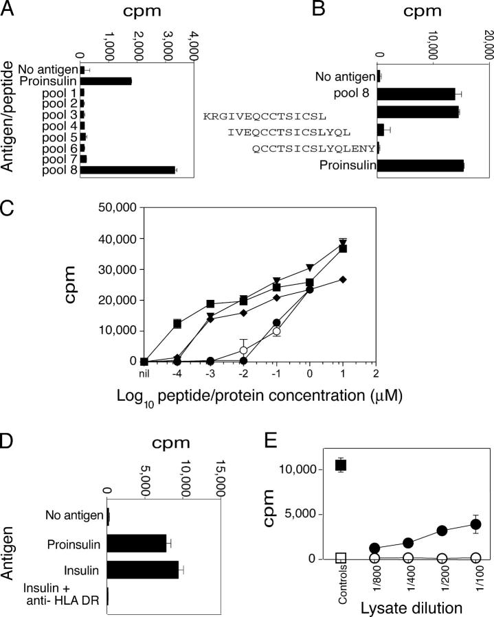
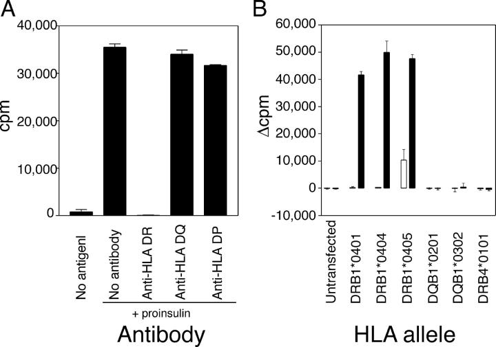
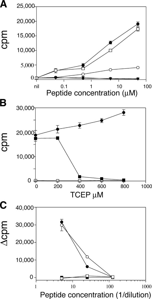
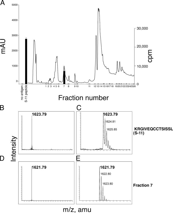
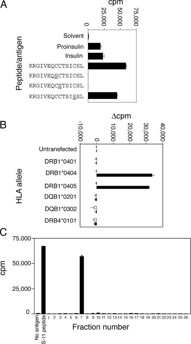
The autoimmune process that destroys the insulin-producing pancreatic beta cells in type 1 diabetes (T1D) is targeted at insulin and its precursor, proinsulin. T cells that recognize the proximal A-chain of human insulin were identified recently in the pancreatic lymph nodes of subjects who had T1D. To investigate the specificity of proinsulin-specific T cells in T1D, we isolated human CD4(+) T cell clones to proinsulin from the blood of a donor who had T1D. The clones recognized a naturally processed, HLA DR4-restricted epitope within the first 13 amino acids of the A-chain (A1-13) of human insulin. T cell recognition was dependent on the formation of a vicinal disulfide bond between adjacent cysteine residues at A6 and A7, which did not alter binding of the peptide to HLA DR4. CD4(+) T cell clones that recognized this epitope were isolated from an HLA DR4(+) child with autoantibodies to insulin, and therefore, at risk for T1D, but not from two healthy HLA DR4(+) donors. We define for the first time a novel posttranslational modification that is required for T cell recognition of the insulin A-chain in T1D.


Figures

Figure 1.
Figure 1.
Identification of the A-chain 1–13 epitope. (A) Preliminary epitope mapping. Peptides, 15-mers shifted by three amino acids, comprising proinsulin, were grouped into eight pools of three to four peptides (5 μg/ml each), or proinsulin (10 μg/ml) were included as indicated. Mean of triplicates ± SEM is shown. (B) Fine epitope mapping. Three peptides from pool 8 were tested separately. (C) Responses to insulin and A-chain peptides. Insulin (open circles) and proinsulin (filled circles) were titrated from 1 μM to 0.1 nM. Peptides KR-A1-13 (filled triangles), A1–13 (filled diamonds), and A1-15 (filled squares) were titrated from 10 μM to 0.1 nM. Other conditions were the same as for A. (D) Responses to insulin. T cell clones were cultured with proinsulin (10 μg/ml), insulin (10 μg/ml), or without antigen (no antigen). Anti-HLA DR mAb (5 μg/ml) was included as indicated. (E) Responses to islet lysate. T cell clones were cultured with dilutions of human islet lysate (filled circles), spleen lysate (open circles), insulin (10 μg/ml; filled square), or without antigen (open square).
Figure 2.
Figure 2.
The response to insulin A1-13 is HLA DR4 restricted. (A) Coarse HLA restriction. Antibodies specific for HLA DR (L243), HLA DQ (SPV-L3), or HLA DP (B7/21) were added to a final concentration of 5 μg/ml. (B) Fine HLA restriction. Insulin-specific T cells were incubated with irradiated, HLA-transfected BLS cells pulsed with 100 μM KR-A1-13 peptide (filled bars) or solvent (open bars). Proliferation of the BLS cells alone after irradiation (1,000–5,000 cpm) was subtracted, and results are expressed as net 3H-thymidine incorporation (Δcpm). Mean ± SEM of triplicates is shown.
Figure 3.
Figure 3.
Oxidized cysteine at A6 and A7, but not antigen processing, is required to elicit responses of T cell clones. (A) Serine for cysteine substitutions. Insulin-specific T cell clones were cultured in the presence of 50—0.05 μM peptide (see Table S2). KR-A1-13 peptide (open squares), S-6 (filled triangles), S-7 (filled diamonds), S-11 (filled circles), or murine KR-A1-13 (open circles). (B) Effect of TCEP on responses to the A1-13 epitope. S-11 peptide (1 μM) was treated with TCEP at the concentrations shown (filled squares). Controls were comprised of either PHA (1.25 μg/ml) and IL-2 (2.5 U/ml; filled circles) or were without antigen (open squares). (C) Effect of APC fixation. HLA-DRB1*0404–transfected BLS cells were fixed cultured with RP-HPLC purified S-11 peptide and T cell clones (filled circles), or with unfixed BLS cells (open circles). Fixed cells with solvent alone (filled squares), or unfixed cells with solvent alone (open squares) were included. The mean ± SEM of triplicate wells is shown.
Figure 4.
Figure 4.
The A1–13 epitope contains a vicinal disulfide bond between cysteine A6 and A7. (A) Isolation of the modified peptide. The absorbance at 214 nm (line) and proliferation of an insulin-specific T cell clone (bars) in response to each fraction (1/400 dilution) is shown. MALDI-QTOF mass spectrometry was used to analyze the parental S-11 peptide at low (B) and high (C) resolution. The active fraction (fraction 7) from the serum-modification experiment at low (D) and high (E) resolution is shown.
Figure 5.
Figure 5.
T cell clones from a donor at high risk for T1D recognize the A1–13 epitope. (A) Isolation of T cell clones. T cell clones were cultured with proinsulin (10 μg/ml), insulin (10 μg/ml), or the KR-A1-13, S-6, S-7, or S-11 peptides (10 μM each), or without antigen. (B) HLA restriction. Irradiated BLS cells that were transfected with the HLA genes shown were pulsed with 100 μM S-11 peptide (filled bars) or solvent alone (open bars). The results are expressed as net cpm proliferation (Δcpm) after subtracting the proliferation of BLS cells alone. (C) Peptide modification in culture medium. Fixed BLS cells that were transfected with HLA DRB1*0404 were cultured with RP-HPLC fractions from Fig. 4. MS analysis of fraction 7 is shown in Fig. 4, D and E. Each point is the mean ± SEM of triplicate wells.

References

    1. Eisenbarth, G.S. 1986. Type I diabetes mellitus. A chronic autoimmune disease. N. Engl. J. Med. 314:1360–1368. - PubMed
    1. Noble, J.A., A.M. Valdes, M. Cook, W. Klitz, G. Thomson, and H.A. Erlich. 1996. The role of HLA class II genes in insulin-dependent diabetes mellitus: molecular analysis of 180 Caucasian, multiplex families. Am. J. Hum. Genet. 59:1134–1148. - PMC - PubMed
    1. Pugliese, A., and G.S. Eisenbarth. 1996. Human type 1 diabetes mellitus: genetic susceptiblity and resistance. Type 1 Diabetes. Molecular, Cellular, and Clinical Immunology. G.S. Eisenbarth and K.J. Lafferty, editors. Oxford University Press, New York. 134–152.
    1. Narendran, P., S.I. Mannering, and L.C. Harrison. 2003. Proinsulin–a pathogenic autoantigen in type 1 diabetes. Autoimmun. Rev. 2:204–210. - PubMed
    1. Pugliese, A., D. Brown, D. Garza, D. Murchison, M. Zeller, M. Redondo, J. Diez, G.S. Eisenbarth, D.D. Patel, and C. Ricordi. 2001. Self-antigen-presenting cells expressing diabetes-associated autoantigens exist in both thymus and peripheral lymphoid organs. J. Clin. Invest. 107:555–564. - PMC - PubMed

Publication types

MeSH terms