. 2005 Nov 7;202(9):1163-9.
doi: 10.1084/jem.20051529. Epub 2005 Oct 31.

Protection from Alzheimer's-like disease in the mouse by genetic ablation of inducible nitric oxide synthase

Affiliations

Protection from Alzheimer's-like disease in the mouse by genetic ablation of inducible nitric oxide synthase

Carl Nathan et al. J Exp Med. .

Abstract

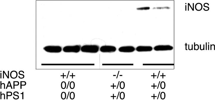
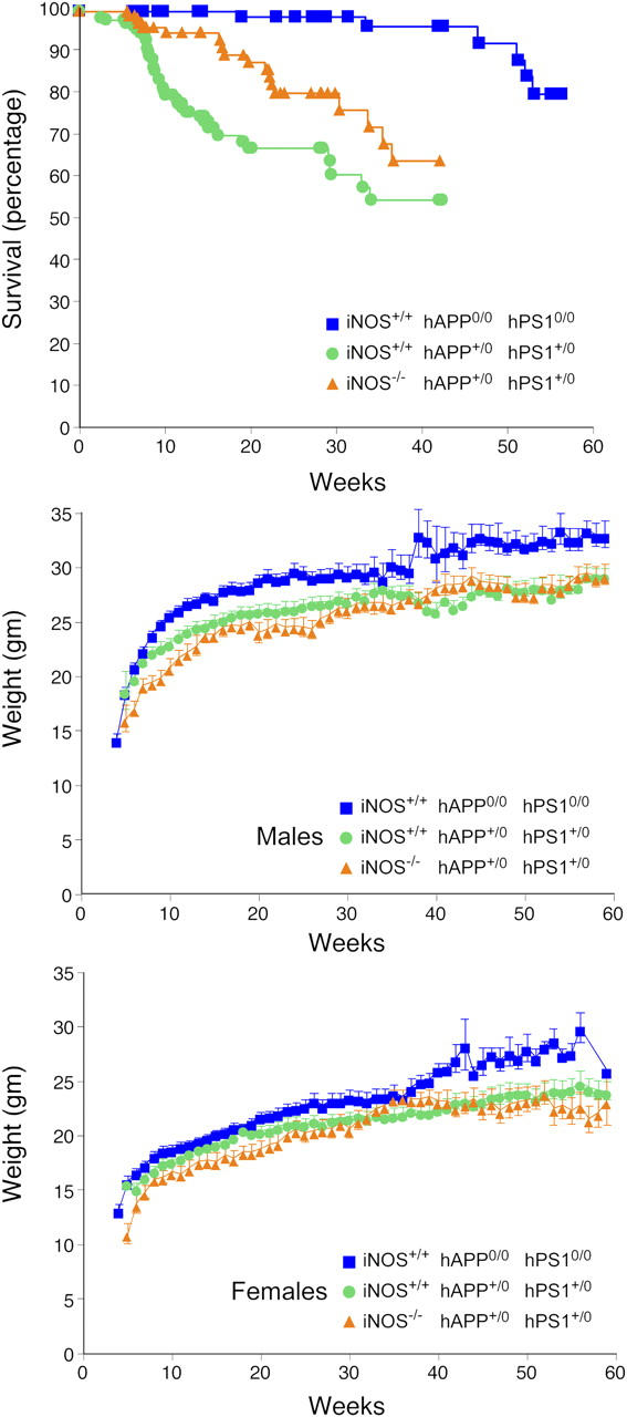
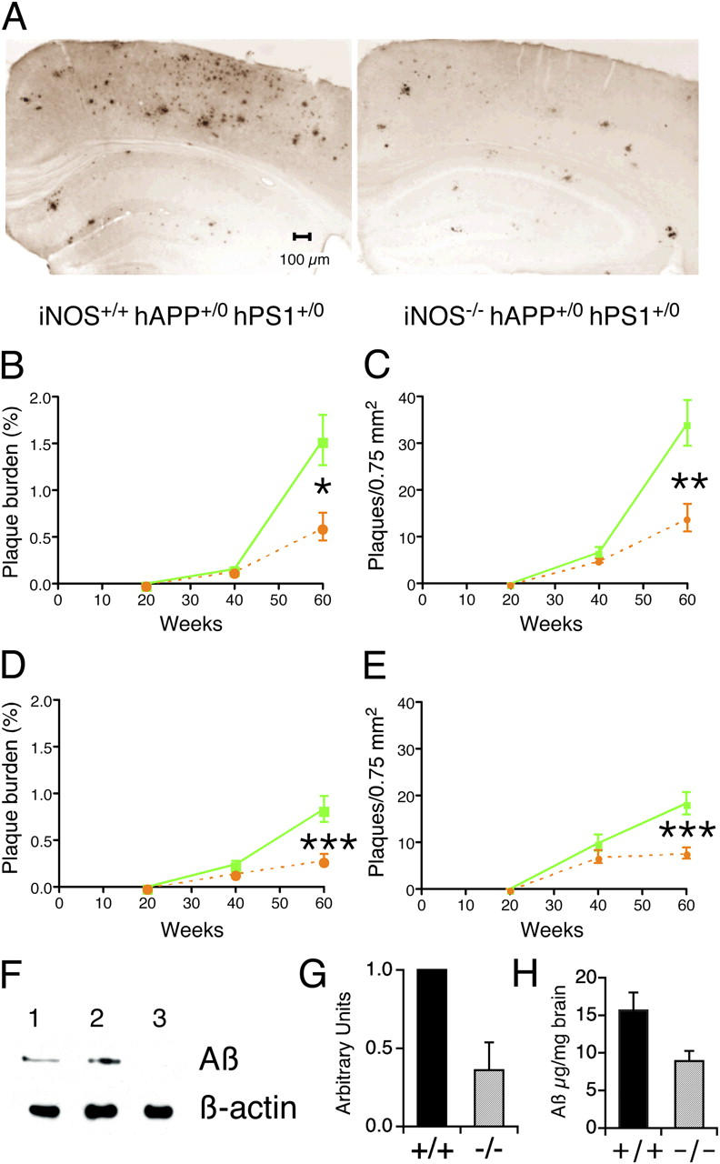
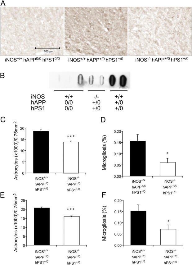
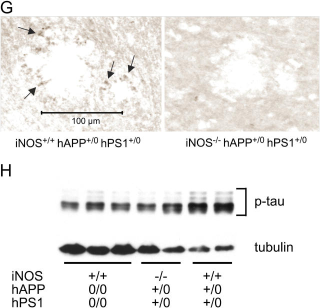
Brains from subjects who have Alzheimer's disease (AD) express inducible nitric oxide synthase (iNOS). We tested the hypothesis that iNOS contributes to AD pathogenesis. Immunoreactive iNOS was detected in brains of mice with AD-like disease resulting from transgenic expression of mutant human beta-amyloid precursor protein (hAPP) and presenilin-1 (hPS1). We bred hAPP-, hPS1-double transgenic mice to be iNOS(+/+) or iNOS(-/-), and compared them with a congenic WT strain. Deficiency of iNOS substantially protected the AD-like mice from premature mortality, cerebral plaque formation, increased beta-amyloid levels, protein tyrosine nitration, astrocytosis, and microgliosis. Thus, iNOS seems to be a major instigator of beta-amyloid deposition and disease progression. Inhibition of iNOS may be a therapeutic option in AD.


Figures

Figure 1.
Figure 1.
Expression of iNOS in brains of 60- to 64-wk-old mice with or without AD-related transgenes, as judged by immunoblot. Each lane is from a separate mouse of the genotypes indicated. Tubulin was immunostained as a loading control.
Figure 2.
Figure 2.
Amelioration of early mortality of AD-transgenic mice by disruption of iNOS alleles without an impact on weight gain. (top, Survival) Individually genotyped mice (iNOS+/+ hAPP0/0 hPS10/0, n = 133; iNOS+/+ hAPP+/0 hPS1+/0, n = 140; iNOS−/− hAPP+/0 hPS1+/0, n = 112) were inspected 5 d per week throughout their lifetime, and their mortality was plotted as a function of age. Differences between any two curves were significant (P < 0.0001; logrank test). (middle) Weight gain in males (iNOS+/+ hAPP0/0 hPS10/0, n = 43; iNOS+/+ hAPP+/0 hPS1+/0, n = 67; iNOS−/− hAPP+/0 hPS1+/0, n = 48). (bottom) Weight gain in females (iNOS+/+ hAPP0/0 hPS10/0, n = 41; iNOS+/+ hAPP+/0 hPS1+/0, n = 50; iNOS−/− hAPP+/0 hPS1+/0, n = 50).
Figure 3.
Figure 3.
Amelioration of late-stage plaque formation and Aβ deposition in AD-transgenic mice by disruption of iNOS alleles. (A) Representative sections from the neocortex and hippocampus of iNOS+/+ hAPP+/0 hPS1+/0 and iNOS−/− hAPP+/0 hPS1+/0 mice immunostained with Aβ (1–42) antibody. (B–E) Plaque burden (percentage of area occupied by Aβ [1–42]-immunoreactive plaques) and plaque numbers per unit area as a function of age in cortex (B and C) and hippocampus (D and E). Means ± SEM for 10 mice per strain per time point. Statistically significant differences are marked (*P < 0.05; **P < 0.01; ***P < 0.003). No immunoreactivity was detected in mice lacking AD transgenes. Solid green line, iNOS+/+ hAPP+/0 hPS1+/0 mice. Dashed orange line, iNOS−/− hAPP+/0 hPS1+/0 mice. (F) Representative immunoblot for brain Aβ that was not extractable in physiologic saline or 0.5% Triton X-100, but was soluble in 6% SDS. Lane 1: iNOS−/− hAPP+/0 hPS1+/0 mice; lane 2: iNOS+/+ hAPP+/0 hPS1+/0 mice; lane 3: iNOS+/+ hAPP0/0 hPS10/0 mice. (G) Densitometry of four blots like that in F, each for different sets of mice, normalized to β-actin. The x axis sets to 1 the ratio of signal intensity for Aβ to that for β-actin in iNOS+/+ hAPP+/0 hPS1+/0 mice (solid bar marked “+/+”); the corresponding ratios for iNOS−/− hAPP+/0 hPS1+/0 mice (hatched bar marked “−/−”) are given as of a proportion of the ratio for the “+/+” mice in each of the same four blots (mean ± SEM). (H) ELISA for Aβ in extracts prepared as in F. Aβ burden is indicated in μg/mg brain weight (mean ± SEM, n = 6) for the two strains with AD-related transgenes whose iNOS alleles are indicated as “+/+” (intact iNOS alleles, black bar) or “−/−“ (disrupted iNOS alleles, gray bar).
Figure 4.
Figure 4.
Amelioration of late-stage nitrosative/oxidative injury, astrogliosis, microgliosis, and phospho-tau accumulation in AD-transgenic mice by disruption of iNOS alleles. (A) Nitrotyrosine immunoreactivity in the cingulate cortex of iNOS+/+ hAPP0/0 hPS10/0, iNOS+/+ hAPP+/0 hPS1+/0, and iNOS−/− hAPP+/0 hPS1+/0 mice. (B) Nitrotyrosine immunoblot. Brain extract proteins (150 μg) were immobilized on a filter with a slot-blot apparatus and were immunoblotted with anti-nitrotyrosine mAb. (C–F) Reduction of GFAP staining indicative of astrocytosis (C and E) and of CD40 staining indicative of microgliosis (D and F) in the cortex (C and D) and hippocampus (E and F) of iNOS−/− hAPP+/0 hPS1+/0 mice. Statistically significant differences are marked (***P < 0.001; *P < 0.05). (G) Reduction of phospho-tau immunoreactivity around plaques in iNOS−/− hAPP+/0 hPS1+/0 mice. Arrows highlight positive staining. (H) Anti–phospho-tau (p-tau) immunoblot with anti-tubulin as a loading control. Each lane is from a separate mouse of the genotypes indicated. See text for quantitative analysis.
Figure 4.
Figure 4.
Amelioration of late-stage nitrosative/oxidative injury, astrogliosis, microgliosis, and phospho-tau accumulation in AD-transgenic mice by disruption of iNOS alleles. (A) Nitrotyrosine immunoreactivity in the cingulate cortex of iNOS+/+ hAPP0/0 hPS10/0, iNOS+/+ hAPP+/0 hPS1+/0, and iNOS−/− hAPP+/0 hPS1+/0 mice. (B) Nitrotyrosine immunoblot. Brain extract proteins (150 μg) were immobilized on a filter with a slot-blot apparatus and were immunoblotted with anti-nitrotyrosine mAb. (C–F) Reduction of GFAP staining indicative of astrocytosis (C and E) and of CD40 staining indicative of microgliosis (D and F) in the cortex (C and D) and hippocampus (E and F) of iNOS−/− hAPP+/0 hPS1+/0 mice. Statistically significant differences are marked (***P < 0.001; *P < 0.05). (G) Reduction of phospho-tau immunoreactivity around plaques in iNOS−/− hAPP+/0 hPS1+/0 mice. Arrows highlight positive staining. (H) Anti–phospho-tau (p-tau) immunoblot with anti-tubulin as a loading control. Each lane is from a separate mouse of the genotypes indicated. See text for quantitative analysis.

References

    1. Rossi, F., and E. Bianchini. 1996. Synergistic induction of nitric oxide by beta-amyloid and cytokines in astrocytes. Biochem. Biophys. Res. Commun. 225:474–478. - PubMed
    1. Barger, S.W., and A.D. Harmon. 1997. Microglial activation by Alzheimer amyloid precursor protein and modulation by apolipoprotein E. Nature. 388:878–881. - PubMed
    1. Ishii, K., F. Muelhauser, U. Liebl, M. Picard, S. Kuhl, B. Penke, T. Bayer, M. Wiessler, M. Hennerici, K. Beyreuther, et al. 2000. Subacute NO generation induced by Alzheimer's beta-amyloid in the living brain: reversal by inhibition of the inducible NO synthase. FASEB J. 14:1485–1489. - PubMed
    1. Smith, M.A., P.L. Richey Harris, L.M. Sayre, J.S. Beckman, and G. Perry. 1997. Widespread peroxynitrite-mediated damage in Alzheimer's disease. J. Neurosci. 17:2653–2657. - PMC - PubMed
    1. Hensley, K., M.L. Maidt, Z. Yu, H. Sang, W.R. Markesbery, and R.A. Floyd. 1998. Electrochemical analysis of protein nitrotyrosine and dityrosine in the Alzheimer brain indicates region-specific accumulation. J. Neurosci. 18:8126–8132. - PMC - PubMed

Publication types

Substances