. 2005 Nov 8;102(45):16426-31.
doi: 10.1073/pnas.0508448102. Epub 2005 Oct 31.

A cAMP-response element binding protein-induced microRNA regulates neuronal morphogenesis

Affiliations

A cAMP-response element binding protein-induced microRNA regulates neuronal morphogenesis

Ngan Vo et al. Proc Natl Acad Sci U S A. .

Erratum in

  • Proc Natl Acad Sci U S A. 2006 Jan 17;103(3):825

Abstract

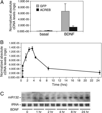
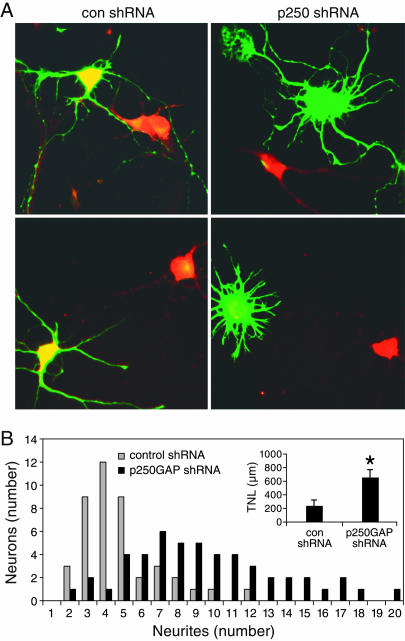
MicroRNAs (miRNAs) regulate cellular fate by controlling the stability or translation of mRNA transcripts. Although the spatial and temporal patterning of miRNA expression is tightly controlled, little is known about signals that induce their expression nor mechanisms of their transcriptional regulation. Furthermore, few miRNA targets have been validated experimentally. The miRNA, miR132, was identified through a genome-wide screen as a target of the transcription factor, cAMP-response element binding protein (CREB). miR132 is enriched in neurons and, like many neuronal CREB targets, is highly induced by neurotrophins. Expression of miR132 in cortical neurons induced neurite outgrowth. Conversely, inhibition of miR132 function attenuated neuronal outgrowth. We provide evidence that miR132 regulates neuronal morphogenesis by decreasing levels of the GTPase-activating protein, p250GAP. These data reveal that a CREB-regulated miRNA regulates neuronal morphogenesis by responding to extrinsic trophic cues.


Figures

Fig. 1.
Fig. 1.
The neuronal miRNA miR132 is regulated by CREB. (A) A diagram indicating the relative positions of genomic signature tags (GSTs, red), CRE motifs (blue), and predicted premiRNA sequences for the miR212 and miR132 cluster. (B) Neocortical neurons were subjected to chromatin immunoprecipitation by using a CREB Ab or IgG control. Real-time PCR was conducted by using primers that interrogate the regions indicated in A.(C) In vivo genomic footprinting was performed in PC12 cells. (Left) Footprint surrounding two consensus CREs upstream of miR212. (Right) Footprint surrounding the single CRE upstream of miR132. The left lanes of each panel indicate dimethyl sulfate-cleaved naked genomic DNA and the right lanes show cleaved DNA from cells treated with dimethyl sulfate in vivo. Arrows denote footprinted bands and asterisks denote hypersensitive sites. (D) Real-time PCR for premiR132 was performed on cDNA from the indicated E18 mouse tissues (hip, hippocampus; cx, neocortex; bs, brainstem; skm, skeletal muscle). The data were normalized to GAPDH cDNA levels.
Fig. 2.
Fig. 2.
miR132 is induced by neurotrophins. (A) Cortical neurons were transfected with dominant negative CREB (ACREB) or vector control (GFP) and treated 36 h later with BDNF for 60 min. RNA was reverse-transcribed and analyzed by real-time PCR with premiR132 primers. The data were normalized to GAPDH cDNA levels. Error is SEM of five to six replicates. (B) Cortical neurons were treated with BDNF for the indicated times. RNA was reverse-transcribed and subjected to real-time PCR with primers specific to premiR132. The data were normalized to GAPDH cDNA levels. Error is SEM of three replicates. (C) Cortical neurons were treated with BDNF for the indicated times. Small molecular weight RNA was isolated and subjected to Northern analysis by using a probe to mature miR132. tRNA levels are shown as a loading control.
Fig. 3.
Fig. 3.
miR132 is transcribed from the stable intron of a cryptic noncoding RNA. (A) A diagram indicating the position of premiR132 and premiR212 and a unique rat noncoding transcript on chromosome 10 (UCSC Genome Center Rn3 assembly). A CpG island and mouse, rat, human, fugu, dog conservation track are indicated. All annotation is from the UCSC Genome Center. The red box in the RT-PCR track indicates a PCR product that interrogates premiR132. (B) Cortical neurons were treated with BDNF or vehicle for 60 min. RNA was reverse-transcribed and analyzed by real-time PCR by using primers interrogating the locations indicated in A. Error is SEM of four replicates.
Fig. 4.
Fig. 4.
Expression of miR132 induces neurite sprouting. (A) Neonatal cortical neurons were transfected with a GFP reporter (green) and cotransfected with vector control, or expression constructs for premiR1-1 or premiR132. Cells were immunostained for the neuronal marker MAP2 (red). (B) Neurons were transfected as in A and analyzed morphometrically. The histogram depicts the distribution of neurons plotted as bins of neurites. The distributions were statistically distinct (P < 0.01; Kruskal-Wallis). (Inset) The average total neurite length (TNL) of miR1-1 (n = 109) and miR132 (n = 137) transfected neurons from four independent experiments. *, P < 0.01 for miR132 vs. miR1-1 (Student's t test).
Fig. 5.
Fig. 5.
Transfection of a 2′O-methyl inhibitor of miR132 markedly attenuated neurite outgrowth. (A) Cortical neurons were transfected with a GFP reporter (green) and cotransfected with empty vector or 2′O-methyl oligoribonucleotide directed against sense or antisense miR132. Cells were immunostained for the neuronal marker MAP2 (red). (B) Neurons were transfected as in A and analyzed morphometrically. The histogram depicts the distribution of neurons plotted as bins of neurites. The distributions were statistically distinct (P < 0.01; Kruskal-Wallis). (Inset) Average total neurite length (TNL) of vector (n = 94), sense (n = 64), and antisense (n = 69) transfected neurons from three independent experiments. *, P < 0.01 for antisense vs. vector or sense (ANOVA, Tukey's posttest).
Fig. 6.
Fig. 6.
p250GAP is a target of miR132. (A) The miR132 target sequence in the p250GAP 3′UTR is conserved across vertebrate evolution. Yellow indicates perfect base pairing, green indicates wobble base pairing, and gray indicates no match. (B) Cortical neurons were cotransfected with GFP-tagged p250GAP and premiRNA132 or premiRNA1-1 expression constructs. LacZ was costransfected as a control. Cell lysates were immunoblotted for GFP and LacZ. -, cells not transfected with p250GAP. (C) Neonatal cortical neurons were transfected with premiR1-1 and premiR132 expression constructs. Cell lysates were immunoblotted for p250GAP or CBP. (D) Cortical neurons were cotransfected with GFP-tagged p250GAP and sense or antisense 2′O-methyl oligos. LacZ was costransfected as a control. Cell lysates were immunoblotted for GFP and LacZ. -, cells not transfected with p250GAP. (E) Neonatal cortical neurons were transfected with sense or antisense 2′O-methyl oligos. Cell lysates were immunoblotted for p250GAP or CBP.
Fig. 7.
Fig. 7.
Down-regulation of p250GAP phenocopies miR132 expression. (A) Cortical neurons were transfected with a GFP reporter (green) and a p250GAP or control shRNA expression construct. Cells were immunostained for p250GAP (red). (B) Neurons were transfected as in A and analyzed morphometrically. The histogram depicts the distribution of neurons plotted as bins of neurites. The distributions were statistically distinct (P < 0.01; Kruskal-Wallis). (Inset) The average total neurite length (TNL) for control shRNA (n = 43) and p250GAP shRNA (n = 50) transfected neurons from two independent experiments. *, P < 0.05 (Student's t test).

References

    1. Lonze, B. E. & Ginty, D. D. (2002) Neuron 35, 605-623. - PubMed
    1. Gonzalez, G. A. & Montminy, M. R. (1989) Cell 59, 675-680. - PubMed
    1. Chrivia, J. C., Kwok, R. P., Lamb, N., Hagiwara, M., Montminy, M. R. & Goodman, R. H. (1993) Nature 365, 855-859. - PubMed
    1. Vo, N. & Goodman, R. H. (2001) J. Biol. Chem. 276, 13505-13508. - PubMed
    1. Conkright, M. D., Canettieri, G., Screaton, R., Guzman, E., Miraglia, L., Hogenesch, J. B. & Montminy, M. (2003) Mol. Cell 12, 413-423. - PubMed

Publication types

Substances

Associated data